薬物依存形成の脳内メカニズムの解明Brain mechanisms in the development of drug addiction
概要Summary
麻薬や覚せい剤などの薬物の摂取は、様々な脳部位で可塑的変化を誘導します。中でも、腹側被蓋野から側坐核および前頭前野に投射するドパミン神経系、いわゆる脳内報酬系(図1)での可塑的変化が薬物依存の形成に重要であると考えられています。これまでの多くの研究から報酬系神経回路での可塑的変化については膨大な知見が得られていますが、報酬系とネットワークを形成する脳幹の神経核での可塑的変化の有無については分かっていません。私たちの研究グループは、電気生理学的・行動薬理学的手法を用いてコカインなどの依存性薬物による脳幹神経核での可塑的変化誘導の可能性を検証し、そのメカニズムを明らかにすることで、薬物依存に対する新たな創薬ターゲットを探索しています。
Use of narcotics and psychostimulants induces plastic changes in various brain regions. Among the plastic changes, those observed in the reward system that is composed of dopaminergic projections from the ventral tegmental area (VTA) to the nucleus accumbens and the prefrontal cortex (Figure 1), are considered to be important for developing drug addiction. Although the plastic changes in the reward system have been extensively studied, the presence of plastic changes in the brainstem nucleus that forms a network with the reward system is still unknown. Our group is studying possible plastic changes induced by cocaine in the brainstem nucleus and investigating their neural mechanisms by electrophysiological and behavioral techniques, thereby discovering a new therapeutic target for drug addiction.
これまでの研究成果Recent results
脳幹の神経核である背外側被蓋核にはグルタミン酸やGABA以外にアセチルコリンを伝達物質として含有するコリン作動性ニューロンが存在し(図1)、腹側被蓋野ドパミンニューロンへ投射しています。背外側被蓋核コリン作動性ニューロンに着目して、可塑的変化の有無を電気生理学的に調べたところ、コカインを慢性摂取したラットの背外側被蓋核コリン作動性ニューロンでは、興奮性シナプス伝達が可塑的に増強していることを見出しました(図2,図3)。また、この可塑的変化の誘導にはNMDA受容体の刺激、一酸化窒素の産生、および、内側前頭前野の活動が必要であることを明らかにしました。この結果は、コカイン摂取により背外側被蓋核コリン作動性ニューロンの活動が上昇し、結果的に、脳内報酬系の活動上昇につながることが、薬物依存の形成に関与する可能性を示唆しています。本研究成果はEuropean Journal of Neuroscience誌で発表し、プレス発表を通して新聞等で紹介されました。
The brainstem laterodorsal tegmental nucleus (LDT) consists of cholinergic, glutamatergic and GABAergic neurons (Figure 1). The LDT projects to dopamine neurons in the VTA. By the use of electrophysiological recordings, we revealed that LDT cholinergic neurons obtained from rats that had been received repeated cocaine administration exhibit plastic enhancement of excitatory synaptic transmission (Figures 2 and 3). We also found that induction of the plastic changes requires stimulation of NMDA receptors, production of nitric oxide and activity of the medial prefrontal cortex. These findings suggest that repeated use of cocaine may enhance activity of LDT cholinergic neurons, which in turn increases activity in the reward system that may be associated with the development of drug addiction. These results have been reported to European Journal of Neuroscience and were released for newspapers and other media at a press conference.
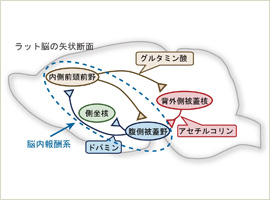

図1 薬物依存の形成に関与すると考えられる神経回路
Figure 1. Neural circuit involved in development of drug addiction.
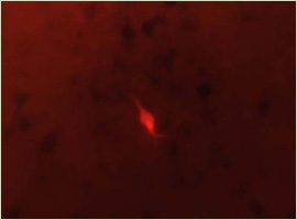
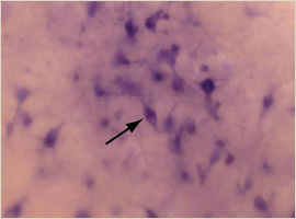
図2 記録した細胞の同定。(左)バイオサイチン染色により記録した細胞が赤色蛍光を発している。(右)背外側被蓋核でのコリン作動性ニューロンのマーカーであるNADPH diaphoraseの染色により、多数のコリン作動性ニューロンが紫色に染まっている。記録した細胞がコリン作動性ニューロンであったことが分かる(矢印)。
Figure 2-Left. Identification of a recorded neuron. Red fluorescent is observed in the cell labelled with biocytin.
Figure 2-Right. Many cholinergic neurons turned purple after staining with NADPH diaphorase, which is a marker for cholinergic neurons in the laterodorsal tegmental nucleus. The labeled cell was identified as a cholinergic neuron (arrow).
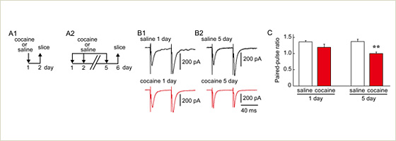
図3 コカイン慢性投与による背外側被蓋核コリン作動性ニューロンでのシナプス可塑性。(A)薬物の投与スケジュール。(B)興奮性シナプス後電流の波形の例。(C)コカインを5日間投与したラットの背外側被蓋核コリン作動性ニューロンでは伝達物質の放出確率の指標であるPaired-pulse ratioが優位に減弱していた。
Figure 3. Synaptic plasticity of cholinergic neurons in the laterodorsal tegmental nucleus following repeated cocaine administration. A1 and A2: experimental schedules, B1 and B2: example traces of excitatory postsynaptic currents, C: The paired-pulse ratio, an index for neurotransmitter release probability, was significantly reduced in cholinergic neurons obtained from rats that had received cocaine for 5 days.