
Research
2. Metabolic pathways of long-chain bases and fatty acid α-oxidation
Biomolecules generally possess not only biosynthetic pathways but also degradative pathways, and homeostasis is maintained through the balance between these processes. One might imagine a system in which unnecessary molecules are simply eliminated by excretion; however, in reality, organisms recover energy through degradation and reuse the components for the synthesis of other biomolecules. In this way, living systems operate through highly efficient and waste-free mechanisms. This principle also applies to ceramides. Ceramides are degraded by ceramidases into fatty acids and long-chain bases. Fatty acids are converted into acyl-CoA for activation, after which they are used for energy production via fatty acid β-oxidation and serve as structural components of lipids. In contrast, long-chain bases are largely limited to reuse in sphingolipid synthesis if left unchanged. Therefore, a degradation pathway exists that converts long-chain bases into fatty acids/acyl-CoA. In this pathway, long-chain bases are first converted into long-chain base 1-phosphates. For example, when the long-chain base is sphingosine, sphingosine 1-phosphate (S1P) is produced. Although S1P is now widely recognized as a lipid mediator, it is thought that during evolution it originally arose as a metabolic product of sphingosine in the long-chain base degradation pathway. S1P acquired its function as a lipid mediator relatively recently in evolution (from chordates onward). In contrast, long-chain base 1-phosphates have served as metabolic intermediates in the long-chain base degradation pathway from evolutionarily ancient times, from yeast to mammals.
Research on S1P as a lipid mediator, as well as on its synthesis and degradation, was actively pursued from the late 1990s to the 2000s. This led to the identification of S1P receptors, enzymes responsible for S1P synthesis (sphingosine kinases) and degradation (S1P lyase and phosphatases), as well as the elucidation of its physiological significance as a lipid mediator through analyses of knockout (KO) mice. These advances culminated in clinical applications, such as the development of a therapeutic agent for multiple sclerosis (fingolimod). In contrast, from the perspective of the degradation pathway, it has been known since the 1960s that long-chain base 1-phosphates undergo cleavage to form long-chain aldehydes, which are subsequently metabolized via acyl-CoAs. However, many details remained unclear, including the genes involved in the pathway, the types and order of reactions, and even, for certain long-chain bases, the metabolic intermediates and final acyl-CoA products. Moreover, the relationship between abnormalities in this pathway and disease had not been elucidated. During our time in the former Laboratory of Biomembrane and Biofunctional Chemistry, we conducted studies focusing on the synthesis, degradation, and release of S1P/long-chain base 1-phosphates as well as fingolimod phosphate (the active form of fingolimod). Subsequently, in 2008, with the start of the Laboratory of Biochemistry, we shifted our focus to the previously unclear degradation steps downstream of long-chain aldehydes in long-chain base degradation pathways and elucidated most of these pathways, along with the pathologies caused by defects in this process.
S1P as a lipid mediator
S1P is a lipid mediator that is present in plasma at concentrations of several hundred nanomolar. S1P acts as a ligand for S1P receptors located on the plasma membrane and induces various cellular responses, including regulation of cell migration, actin cytoskeleton organization, cell proliferation, and adhesion junction formation1). Five S1P receptors, S1PR1 through S1PR5, have been identified, among which S1PR1 is the most physiologically important. These receptors are G protein-coupled receptors, each transducing intracellular signals through specific G proteins (Figure 5).
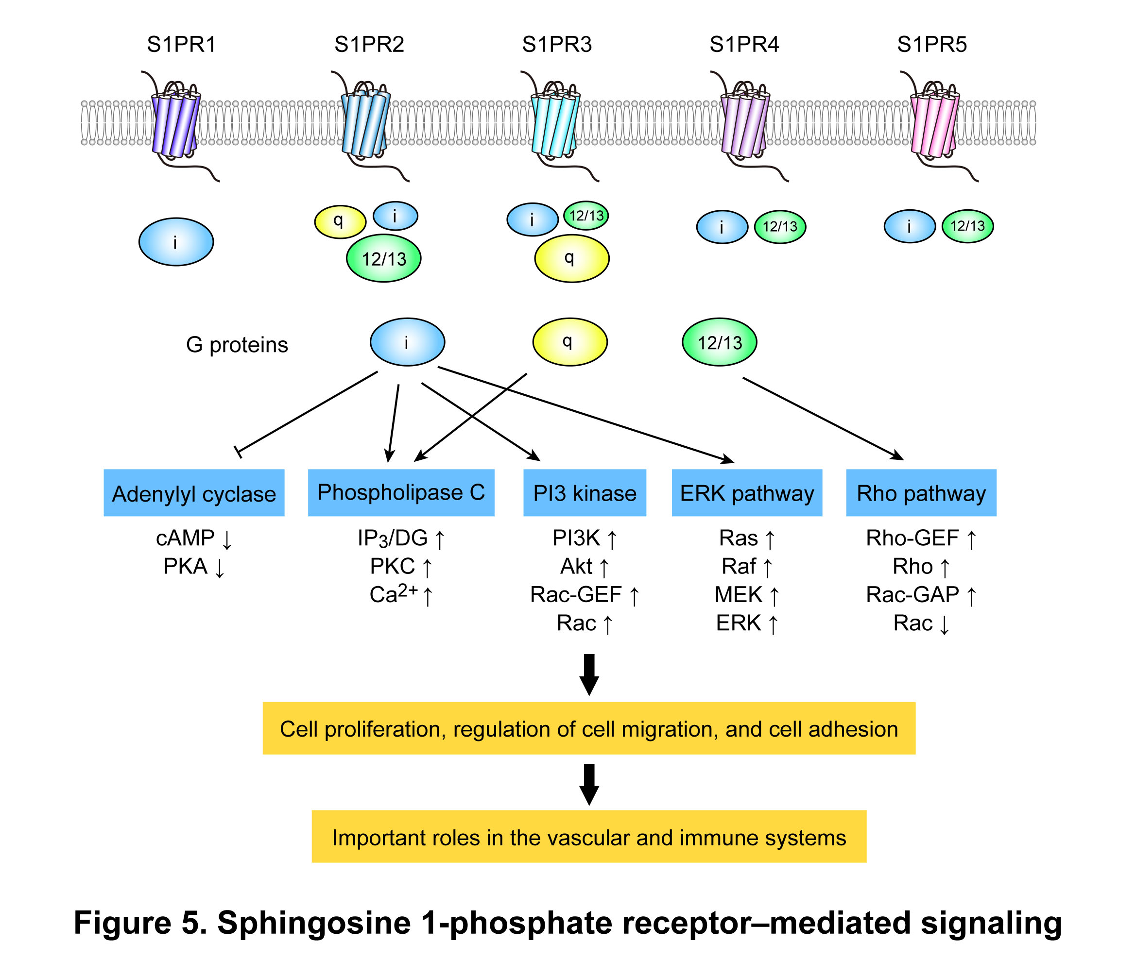
Because S1P is abundant in plasma, its effects on vascular endothelial cells and lymphocytes in contact with plasma are physiologically important. In the vascular system, S1P plays an essential role in vascular formation during embryogenesis, whereas in the immune system, it is critical for the egress of lymphocytes from the thymus and secondary lymphoid organs. After maturing in the thymus, T lymphocytes enter the bloodstream and circulate between the blood and secondary lymphoid organs (Figure 6). Naïve T lymphocytes migrate into secondary lymphoid organs and, if they do not encounter antigen, return to the bloodstream. When they recognize antigens presented by antigen-presenting cells, they differentiate into effector cells, proliferate, and then return to the bloodstream. Egress from the thymus and secondary lymphoid organs is mainly mediated by S1P and S1PR12). S1PR1 on lymphocytes induces chemotaxis in response to S1P concentration gradients between the thymus–plasma and the secondary lymphoid organs–lymph–plasma. On endothelial cells, S1PR1 regulates intercellular adhesion. If S1PR1 on lymphocytes is absent or the S1P concentration gradient is disrupted, T lymphocytes fail to exit from the thymus or secondary lymphoid organs, resulting in a reduction of circulating T lymphocytes and consequent immunosuppression. Fingolimod (originally developed as FTY720), an immunomodulatory drug used to treat multiple sclerosis, is phosphorylated in vivo to form fingolimod phosphate, which acts as a ligand for S1PR1. It downregulates S1PR1 more potently than S1P by promoting its degradation in lysosomes without recycling to the cell surface, thereby inducing immunosuppression.
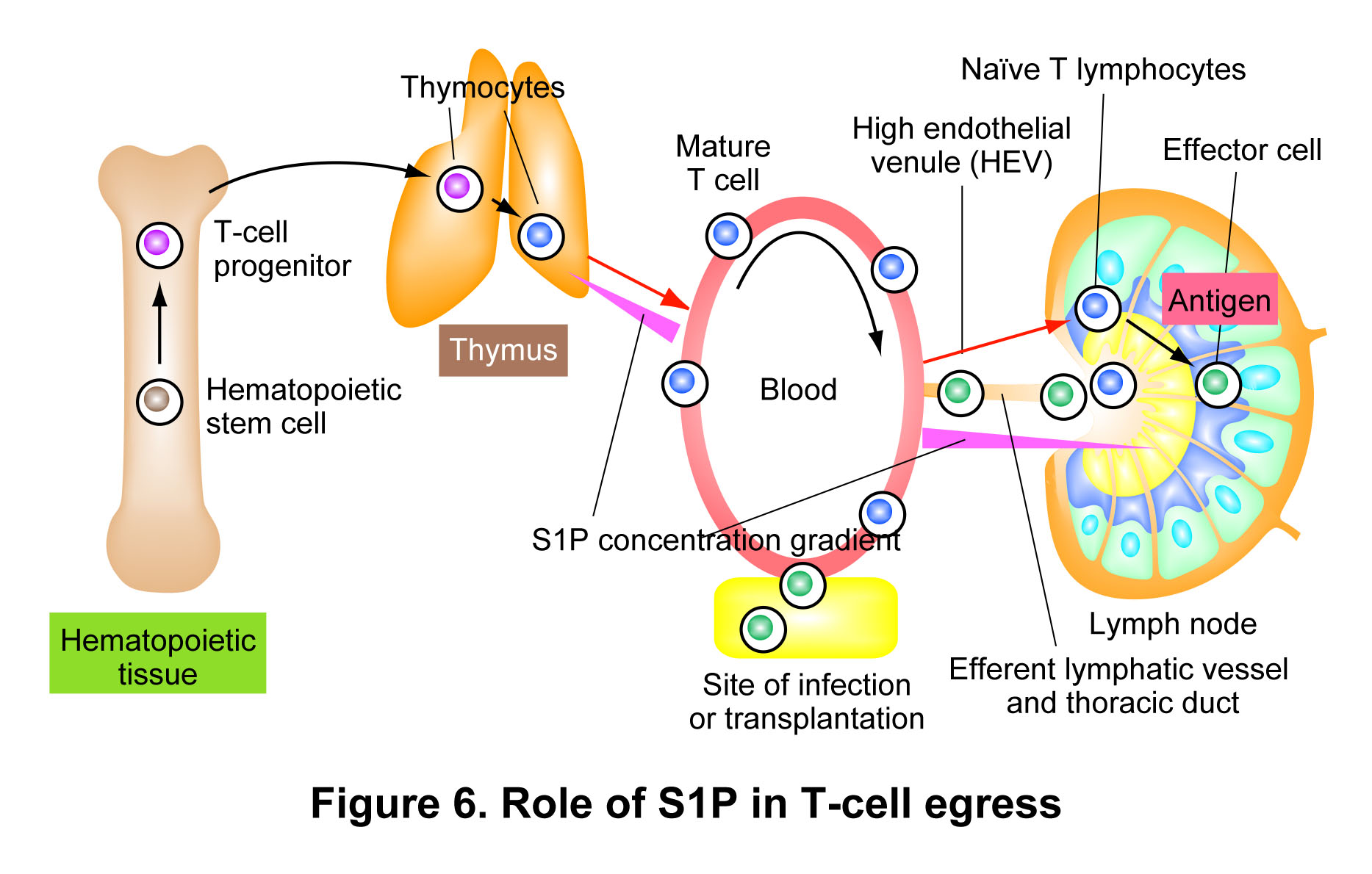
As described above, maintaining the S1P concentration gradient between tissues and plasma is critically important. In general, S1P levels in tissues are kept low because the activities of the degrading enzymes S1P lyase and S1P phosphatase predominate over that of the synthetic enzyme, sphingosine kinase. In contrast, the concentration of S1P in plasma is determined by the balance between its supply from erythrocytes and endothelial cells and its uptake by cells exposed to plasma and its degradation by cell surface phosphatases. To date, we have identified the following aspects of the metabolic enzymes of S1P/long-chain bases and fingolimod and fingolimod phosphate, as well as their dynamics.
- We identified SGPP2 (also known as SPP2) as a human S1P phosphatase gene and characterized the enzymatic properties of its gene product3).
- We demonstrated that both S1P phosphatase and S1P lyase are localized to the endoplasmic reticulum, with their catalytic domains positioned on opposite sides of the ER membrane, facing the luminal and cytosolic sides, respectively4, 5).
- We showed that erythrocytes lack S1P-degrading enzymes and therefore accumulate S1P at high levels, functioning as a major source of plasma S1P6).
- We found that platelets, rather than erythrocytes, serve as the primary source of fingolimod phosphate in plasma7).
- We demonstrated that fingolimod phosphate is dephosphorylated and inactivated by the phospholipid phosphatases PLPPs (also known as LPPs) on the plasma membrane of vascular endothelial cells8).
- We found that mouse sphingosine kinase 1 transcriptional variants (SPHK1a and SPHK1b) differ markedly in their enzymatic properties, including subcellular localization, oligomerization state, post-translational modification, and stability9).
- We demonstrated that most extracellular S1P is dephosphorylated by the phospholipid phosphatases PLPPs on the cell surface and subsequently taken up into cells; however, a fraction is directly taken up as S1P, particularly in erythrocytes10). We further showed that this uptake involves SPNS2 and MFSD2B, which were previously characterized as S1P exporters, but function as channel-type transporters capable of bidirectional transport across the plasma membrane10).
- We found that extracellular long-chain bases are rapidly taken up into cells in a process dependent on acyl-CoA synthetases (Faa1 and Faa4 in yeast, and ACSLs in mammals)11). In mammals, some acyl-CoA synthetases were originally identified as fatty acid transporters. This observation suggests that acyl-CoA synthetases involved in long-chain base uptake may also function directly as transporters for long-chain bases.
- We identified Rsb1 as a transporter responsible for exporting long-chain bases from cells to the extracellular space in yeast12).
- We found that the yeast sphingosine kinase Lcb4 undergoes multiple post-translational modifications (palmitoylation, phosphorylation, and ubiquitination) and elucidated the mechanisms by which these modifications regulate its subcellular localization and stability13–15).
Long-chain base degradation pathway and S1P as a metabolic intermediate of sphingosine
S1P functions as a lipid mediator in the extracellular space, whereas it is produced intracellularly. Although most cells produce S1P, its release into the extracellular space is limited to specific cell types such as blood cells and vascular/lymphatic endothelial cells. In other cells (and likely even in endothelial cells that release S1P), the majority of S1P is metabolized intracellularly without being secreted. Although it has been proposed to function as a second messenger, it is primarily produced as a metabolic intermediate. The ceramide moiety of sphingolipids is converted into fatty acids and long-chain bases, such as sphingosine, by hydrolysis mediated by ceramidases. Long-chain bases, such as sphingosine, are either reused for sphingolipid synthesis through reacylation to form ceramides (the salvage pathway) or converted into acyl-CoAs (e.g., palmitoyl-CoA in the case of an 18-carbon sphingosine) via long-chain base 1-phosphates, including S1P (Figure 7). Acyl-CoAs are then used either directly in lipid metabolism (primarily for glycerolipids and partly for other lipid classes) or after fatty acid elongation and desaturation, or in fatty acid β-oxidation. The long-chain base degradation pathway is the sole pathway that converts the long-chain base moiety of sphingolipids into acyl-CoAs. Therefore, this pathway plays an essential role in maintaining sphingolipid homeostasis, and its disruption impairs normal cellular functions. Indeed, mutations in the SGPL1 gene, which encodes S1P lyase that catalyzes the first irreversible step in long-chain base metabolism, cause RENI syndrome, a hereditary disorder characterized by multisystem abnormalities. Consistently, Sgpl1 knockout (KO) mice exhibit metabolic abnormalities in the liver as well as morphological defects in the lung, heart, ureter, and bone, and display a markedly shortened lifespan of approximately one month.
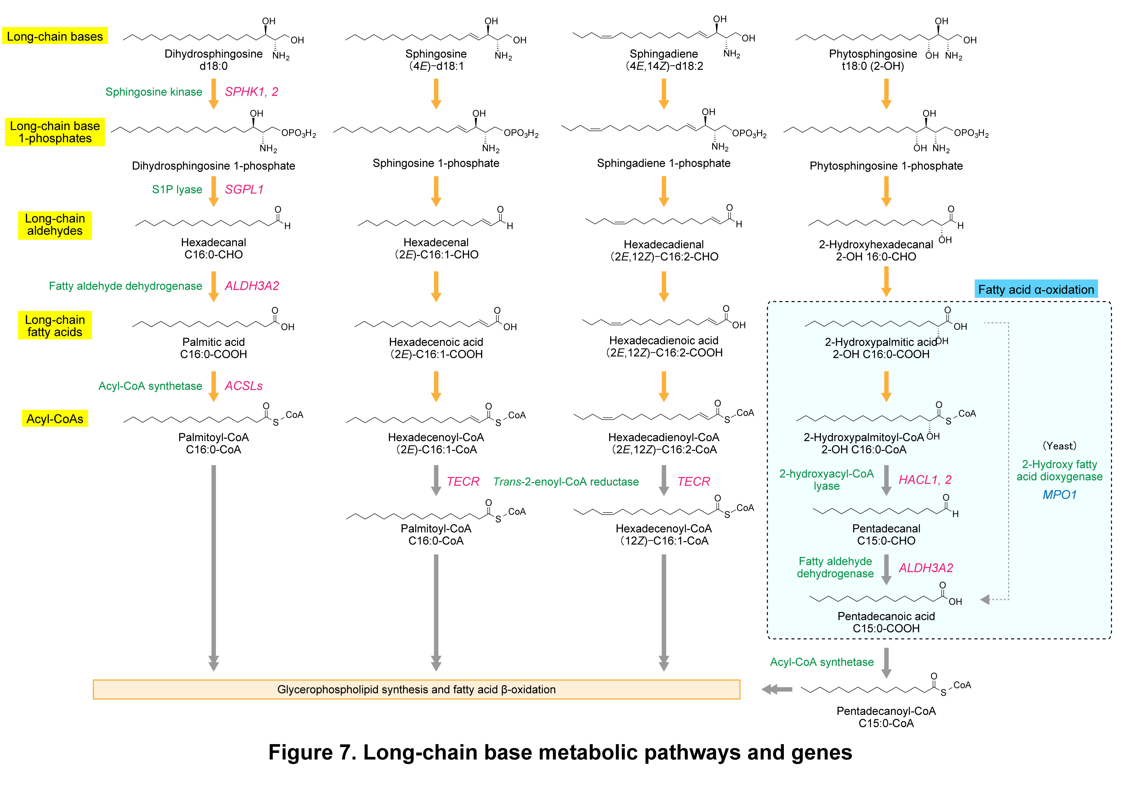
In the sphingosine degradation pathway, the metabolic steps downstream of trans-2-hexadecenal, the product of S1P lyase, as well as the genes involved, remained unclear for a long time. Our laboratory addressed this issue and elucidated the complete long-chain base degradation pathway, including sphingosine, dihydrosphingosine, phytosphingosine, and sphingadiene (Figure 7)16, 17). In the long-chain base degradation pathway, common steps are conserved regardless of long-chain base type, namely phosphorylation (generation of long-chain base 1-phosphates), cleavage (generation of long-chain aldehydes), oxidation (generation of long-chain fatty acids), and CoA conjugation (generation of acyl-CoAs) (Figure 7, orange arrows). Our laboratory identified the enzymes responsible for the latter two steps of these common reactions: fatty aldehyde dehydrogenases, which catalyze the oxidation step (ALDH3A2 in mammals and Hfd1 in yeast), and acyl-CoA synthetases, which catalyze CoA addition (primarily ACSLs in mammals and Faa1 and Faa4 in yeast)18, 19). In the degradation of long-chain bases with a carbon number of n, cleavage by SGPL1 produces an (n−2)-carbon long-chain aldehyde and a C2 compound, ethanolamine phosphate. The resulting (n−2)-carbon aldehyde is subsequently converted into the corresponding (n−2)-carbon acyl-CoA. In the case of phytosphingosine, however, an α-oxidation step is involved, resulting in the production of an (n−3)-carbon acyl-CoA. The metabolism of C18 long-chain bases can be summarized as follows. Long-chain bases are designated according to the number of hydroxyl groups, carbon chain length, and degree of unsaturation; for example, dihydrosphingosine with the composition C18:0 is denoted as d18:0. The prefix “d” indicates two hydroxyl groups (di), whereas “t” denotes long-chain bases containing three hydroxyl groups, such as phytosphingosine (tri). The simplest long-chain base, dihydrosphingosine (d18:0), is converted into palmitoyl-CoA via this pathway. Sphingosine (d18:1), which contains a trans double bond between C4–5, is likewise converted into palmitoyl-CoA; however, this conversion requires saturation of the double bond. Our laboratory demonstrated that trans-2-enoyl-CoA reductase (TECR) catalyzes the conversion of trans-2-hexadecenoyl-CoA to palmitoyl-CoA20). Similarly, sphingadiene (d18:2) undergoes saturation of the trans double bond between C4–5, yielding cis-12-hexadecenoyl-CoA21).
The fatty aldehyde dehydrogenase gene ALDH3A2, which is involved in long-chain base metabolism, is the causative gene for Sjögren–Larsson syndrome (SLS) (see also “4. Ceramide-mediated skin barrier formation”). SLS is an inherited disorder characterized by ichthyosis, intellectual disability, and spastic diplegia, and the accumulation of fatty aldehydes, which are substrates of ALDH3A2, is thought to contribute to its pathogenesis. Although the specific fatty aldehyde that acts as the primary causative molecule remains unclear, we propose that trans-2-hexadecenal derived from sphingosine contributes to the development of ichthyotic symptoms in SLS.
ALDH3A2 plays a central role in the metabolism of long-chain aldehydes derived from long-chain bases; however, the long-chain base degradation pathway is not completely blocked even in cells lacking ALDH3A2. This is because other members of the ALDH3 family can compensate for the loss of ALDH3A2. Our laboratory demonstrated that ALDH3B1, ALDH3B2 (whose gene is pseudogenized in humans and therefore does not produce a functional protein), and ALDH3B3 exhibit high enzymatic activity toward long-chain aldehydes, including trans-2-hexadecenal, similar to ALDH3A222, 23). These fatty aldehyde dehydrogenases share similar substrate specificities but differ in their subcellular localization (ALDH3A2 in the endoplasmic reticulum, ALDH3B1 and ALDH3B3 at the plasma membrane, and ALDH3B2 in lipid droplets)22, 23). Among fatty aldehyde dehydrogenases, ALDH3A2 is the major contributor to long-chain base metabolism. This is likely because the reactions involved in long-chain base degradation occur in the endoplasmic reticulum, where ALDH3A2 is localized.
Epidermal analysis of Aldh3a2 KO mice revealed no significant changes in fatty aldehyde dehydrogenase activity compared with wild-type mice, with no defects in skin barrier formation or changes in transepidermal water loss24). This apparent lack of phenotype is attributable to functional compensation by Aldh3b2, which is present specifically in mice, as evidenced by Aldh3a2 Aldh3b2 double KO mice exhibiting defects in skin barrier function25). In contrast, loss of Aldh3a2 in the brain reduces fatty aldehyde dehydrogenase activity to approximately 30% of that in wild-type mice and results in mild motor dysfunction26). In the brains of Aldh3a2 KO mice, levels of 2-hydroxygalactosylceramide, a sphingolipid important for myelin maintenance, are decreased. This decrease is likely due to the accumulation of trans-2-hexadecenal in Aldh3a2 KO mice, which is accommodated in the substrate-binding pocket of the fatty acid 2-hydroxylase FA2H and covalently modifies active-site histidine residues26).
Fatty acid α-oxidation
Among long-chain bases in ceramides, phytosphingosine, which possesses a hydroxyl group at C-4, is found in mammals only in epithelial tissues such as the epidermis, esophagus, stomach, small intestine, large intestine, and kidney27), whereas it represents the major long-chain base in yeast. Our laboratory found that phytosphingosine, unlike sphingosine or dihydrosphingosine, is metabolized into odd-chain fatty acids (e.g., pentadecanoic acid from t18:0 phytosphingosine) (Figure 7)28). The initial steps of phytosphingosine metabolism are similar to those of sphingosine and dihydrosphingosine, proceeding sequentially through long-chain base 1-phosphate (phytosphingosine 1-phosphate), long-chain aldehyde (2-hydroxyhexadecanal), and long-chain fatty acid (2-hydroxypalmitic acid). Because phytosphingosine contains a hydroxyl group at C-4, this process generates a fatty acid with a hydroxyl group at C-2 (2-hydroxypalmitic acid). The presence of this hydroxyl group determines the subsequent metabolic steps, as 2-hydroxypalmitic acid undergoes α-oxidation, resulting in both the shortening of the carbon chain by one carbon and the removal of the hydroxyl group. Our laboratory identified MPO1 as the gene responsible for this α-oxidation step in the phytosphingosine degradation pathway in yeast28). Mpo1 belongs to the DUF962 protein family (DUF: Domain of Unknown Function), which is conserved from prokaryotes to lower eukaryotes. Although more than 3,000 proteins have been identified in this family, Mpo1 was the first member for which a biological function was elucidated. Subsequent analyses revealed that Mpo1 is a novel dioxygenase that utilizes an iron ion as a cofactor and converts 2-hydroxy fatty acids into non-hydroxylated fatty acids with one fewer carbon atom29). The activity of Mpo1 requires three highly conserved histidine residues that coordinate the iron ion30). In addition to long-chain 2-hydroxy fatty acids derived from the phytosphingosine degradation pathway, Mpo1 also utilizes 2-hydroxy fatty acids generated through general fatty acid metabolism, including very-long-chain species30). Deletion of the MPO1 gene does not affect yeast growth under normal growth conditions but causes growth retardation under conditions of endoplasmic reticulum stress or carbon starvation30).
Many of the metabolic pathways and genes involved in long-chain base metabolism are conserved from yeast to mammals. However, fatty acid α-oxidation in the phytosphingosine metabolic pathway differs between yeast and mammals (Figure 7). Mammals lack Mpo1 homologs; instead, our laboratory found that 2-hydroxypalmitic acid is converted into 2-hydroxypalmitoyl-CoA and cleaved mainly by the 2-hydroxyacyl-CoA lyase HACL2 (and to a lesser extent HACL1) to form pentadecanal, which is then converted into pentadecanoic acid by the fatty aldehyde dehydrogenase ALDH3A231). HACL2 is a 2-hydroxyacyl-CoA lyase identified by our laboratory and localized in the endoplasmic reticulum31). Fatty acid α-oxidation is known to occur in peroxisomes; however, our findings revealed for the first time that it also occurs in the endoplasmic reticulum. Although odd-chain fatty acids are present in vivo at low levels, they were previously considered to be generated by fatty acid synthase using propionyl-ACP in place of acetyl-ACP. Our results demonstrate that, in addition to this mechanism, odd-chain fatty acids are produced via α-oxidation of 2-hydroxy fatty acids derived from the phytosphingosine degradation pathway and from fatty acid 2-hydroxylation. Indeed, in the brains of Hacl2 KO mice, levels of odd-chain monohexosylceramides (most of which are galactosylceramides) are reduced to approximately one-quarter of those in wild-type mice, while odd-chain ceramides in the stomach are reduced by approximately half32).
The substrates for fatty acid α-oxidation are not limited to 2-hydroxy fatty acids but also include branched fatty acids methylated at C-3. Our laboratory demonstrated that the two 2-hydroxyacyl-CoA lyase isozymes, HACL1 and HACL2, are redundantly involved in the metabolism of both 2-hydroxy fatty acids and 3-methyl fatty acids, whereas HACL1 primarily catalyzes the α-oxidation of 3-methyl fatty acids and HACL2 primarily catalyzes that of 2-hydroxy fatty acids32). The physiological role of fatty acid α-oxidation is not the production of odd-chain fatty acids, but rather the conversion of 2-hydroxy fatty acids and 3-methyl fatty acids, which cannot undergo β-oxidation, into metabolizable non-hydroxylated fatty acids and 2-methyl fatty acids, respectively. In general, biomolecules maintain homeostasis through a balance between synthesis and degradation, and disruption of this balance leads to various cellular dysfunctions and diseases. The degradation pathways of long-chain bases elucidated by our laboratory provide a molecular framework for understanding sphingolipid homeostasis from the perspective of degradation.
References
- Kihara A, Mitsutake S, Mizutani Y, Igarashi Y. (2007) Metabolism and biological functions of two phosphorylated sphingolipids, sphingosine 1-phosphate and ceramide 1-phosphate. Prog Lipid Res, 46, 126–144, Review.
- Kihara A, Igarashi Y. (2008) Production and release of sphingosine 1-phosphate and the phosphorylated form of the immunomodulator FTY720. Biochim Biophys Acta, 1781, 496–502, Review.
- Ogawa C, Kihara A, Gokoh M, Igarashi Y. (2003) Identification and characterization of a novel human sphingosine-1-phosphate phosphohydrolase, hSPP2. J Biol Chem, 278, 1268–1272.
- Kihara A, Sano T, Iwaki S, Igarashi Y. (2003) Transmembrane topology of sphingoid long-chain base-1-phosphate phosphatase, Lcb3p. Genes Cells, 8, 525–535.
- Ikeda M, Kihara A, Igarashi Y. (2004) Sphingosine-1-phosphate lyase SPL is an endoplasmic reticulum-resident, integral membrane protein with the pyridoxal 5'-phosphate binding domain exposed to the cytosol. Biochem Biophys Res Commun, 325, 338–343.
- Ito K, Anada Y, Tani M, Ikeda M, Sano T, Kihara A, Igarashi Y. (2007) Lack of sphingosine 1-phosphate-degrading enzymes in erythrocytes. Biochem Biophys Res Commun, 357, 212–217.
- Anada Y, Igarashi Y, Kihara A. (2007) The immunomodulator FTY720 is phosphorylated and released from platelets. Eur J Pharmacol, 568, 106–111.
- Yamanaka M, Anada Y, Igarashi Y, Kihara A. (2008) A splicing isoform of LPP1, LPP1a, exhibits high phosphatase activity toward FTY720 phosphate. Biochem Biophys Res Commun, 375, 675–679.
- Kihara A, Anada Y, Igarashi Y. (2006) Mouse sphingosine kinase isoforms SPHK1a and SPHK1b differ in enzymatic traits including stability, localization, modification, and oligomerization. J Biol Chem, 281, 4532–4539.
- Goto H, Miyamoto M, Kihara A. (2021) Direct uptake of sphingosine-1-phosphate independent of phospholipid phosphatases. J Biol Chem, 296, 100605.
- Narita T, Naganuma T, Sase Y, Kihara A. (2016) Long-chain bases of sphingolipids are transported into cells via the acyl-CoA synthetases. Sci Rep, 6, 25469.
- Kihara A, Igarashi Y. (2002) Identification and characterization of a Saccharomyces cerevisiae gene, RSB1, involved in sphingoid long-chain base release. J Biol Chem, 277, 30048–30054.
- Kihara A, Kurotsu F, Sano T, Iwaki S, Igarashi Y. (2005) Long-chain base kinase Lcb4 Is anchored to the membrane through its palmitoylation by Akr1. Mol Cell Biol, 25, 9189–9197.
- Iwaki S, Kihara A, Sano T, Igarashi Y. (2005) Phosphorylation by Pho85 cyclin-dependent kinase acts as a signal for the down-regulation of the yeast sphingoid long-chain base kinase Lcb4 during the stationary phase. J Biol Chem, 280, 6520–6527.
- Sano T, Kihara A, Kurotsu F, Iwaki S, Igarashi Y. (2005) Regulation of the sphingoid long-chain base kinase Lcb4p by ergosterol and heme: studies in phytosphingosine-resistant mutants. J Biol Chem, 280, 36674–36682.
- Kihara A. (2015) Sphingolipid metabolism via sphingosine 1-phosphate and its role in physiology pathology, and nutrition. in Bioactive lipid mediators: current reviews and protocols (Yokomizo, T., and Murakami, M. eds.), Springer Japan, Tokyo, Review.
- Kihara A. (2016) Synthesis and degradation pathways, functions, and pathology of ceramides and epidermal acylceramides. Prog Lipid Res, 63, 50–69.
- Nakahara K, Ohkuni A, Kitamura T, Abe K, Naganuma T, Ohno Y, Zoeller R A, Kihara A. (2012) The Sjögren-Larsson syndrome gene encodes a hexadecenal dehydrogenase of the sphingosine 1-phosphate degradation pathway. Mol Cell, 46, 461–471.
- Ohkuni A, Ohno Y, Kihara A. (2013) Identification of acyl-CoA synthetases involved in the mammalian sphingosine 1-phosphate metabolic pathway. Biochem Biophys Res Commun, 442, 195–201.
- Wakashima T, Abe K, Kihara A. (2014) Dual functions of the trans-2-enoyl-CoA reductase TER in the sphingosine 1-phosphate metabolic pathway and in fatty acid elongation. J Biol Chem, 289, 24736–24748.
- Jojima K, Kihara A. (2023) Metabolism of sphingadiene and characterization of the sphingadiene-producing enzyme FADS3. Biochim Biophys Acta Mol Cell Biol Lipids, 1868, 159335.
- Kitamura T, Naganuma T, Abe K, Nakahara K, Ohno Y, Kihara A. (2013) Substrate specificity, plasma membrane localization, and lipid modification of the aldehyde dehydrogenase ALDH3B1. Biochim Biophys Acta, 1831, 1395–1401.
- Kitamura T, Takagi S, Naganuma T, Kihara A. (2015) Mouse aldehyde dehydrogenase ALDH3B2 is localized to lipid droplets via two C-terminal tryptophan residues and lipid modification. Biochem J, 465, 79–87.
- Naganuma T, Takagi S, Kanetake T, Kitamura T, Hattori S, Miyakawa T, Sassa T, Kihara A. (2016) Disruption of the Sjögren-Larsson syndrome gene Aldh3a2 in mice increases keratinocyte growth and retards skin barrier recovery. J Biol Chem, 291, 11676–11688.
- Nojiri K, Fudetani S, Arai A, Kitamura T, Sassa T, Kihara A. (2021) Impaired skin barrier function due to reduced ω-O-acylceramide levels in a mouse model of Sjögren-Larsson syndrome. Mol Cell Biol, 41, e0035221.
- Kanetake T, Sassa T, Nojiri K, Sawai M, Hattori S, Miyakawa T, Kitamura T, Kihara A. (2019) Neural symptoms in a gene knockout mouse model of Sjögren-Larsson syndrome are associated with a decrease in 2-hydroxygalactosylceramide. FASEB J, 33, 928–941.
- Ota A, Morita H, Naganuma T, Miyamoto M, Jojima K, Nojiri K, Matsuda J, Kihara A. (2023) Bifunctional DEGS2 has higher hydroxylase activity toward substrates with very-long-chain fatty acids in the production of phytosphingosine ceramides. J Biol Chem, 299, 104603.
- Kondo N, Ohno Y, Yamagata M, Obara T, Seki N, Kitamura T, Naganuma T, Kihara A. (2014) Identification of the phytosphingosine metabolic pathway leading to odd-numbered fatty acids. Nat Commun, 5, 5338.
- Seki N, Mori K, Kitamura T, Miyamoto M, Kihara A. (2019) Yeast Mpo1 is a novel dioxygenase that catalyzes the α-oxidation of a 2-hydroxy fatty acid in an Fe2+-dependent manner. Mol Cell Biol, 39, e00428-00418.
- Mori K, Obara T, Seki N, Miyamoto M, Naganuma T, Kitamura T, Kihara A. (2020) Catalytic residues, substrate specificity, and role in carbon starvation of the 2-hydroxy FA dioxygenase Mpo1 in yeast. J Lipid Res, 61, 1104–1114.
- Kitamura T, Seki N, Kihara A. (2017) Phytosphingosine degradation pathway includes fatty acid α-oxidation reactions in the endoplasmic reticulum. Proc Natl Acad Sci USA, 114, E2616–E2623.
- Mori K, Naganuma T, Kihara A. (2023) Role of 2-hydroxy acyl-CoA lyase HACL2 in odd-chain fatty acid production via α-oxidation in vivo. Mol Biol Cell, 34, ar85.