
Research
4. Ceramide-mediated skin barrier formation
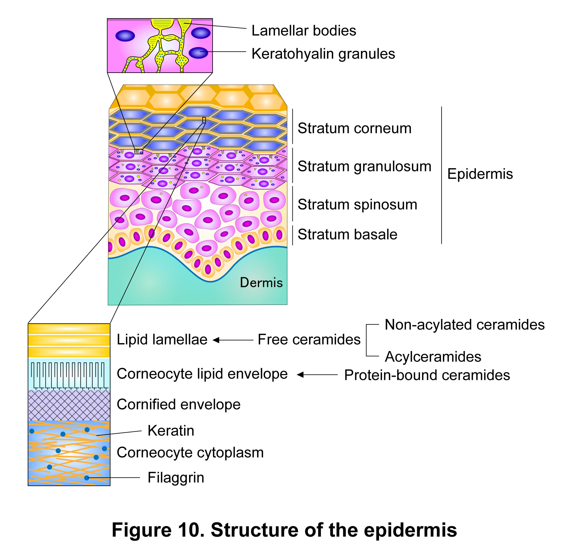
The epidermis is composed of four layers: the stratum basale, stratum spinosum, stratum granulosum, and stratum corneum. Of these, the outermost layer, the stratum corneum, plays the major role in forming the skin permeability barrier (skin barrier; Figure 10). The skin barrier is essential for preventing the entry of pathogens, foreign substances, and allergens that cause infections, inflammation, and allergic diseases, and for retaining water within the body. Therefore, impaired barrier function increases the risk of, or directly contributes to, various skin disorders, including infections, atopic dermatitis, ichthyosis (epidermal differentiation disorders [EDDs]), and xerosis.
Ceramide-containing structures in the stratum corneum
Ceramides constitute two key structures in the stratum corneum that are fundamental to skin barrier formation: the lipid lamellae and the corneocyte lipid envelope (CLE). The lipid lamellae are multilayered lipid structures located in the intercellular spaces of the stratum corneum and exhibit two characteristic periodicities in humans, with a long periodicity phase of approximately 13 nm and a short periodicity phase of approximately 6 nm. The major components of lipid lamellae are ceramides, cholesterol, and free fatty acids. Previous reviews have described these three lipid classes as being present at approximately equimolar levels. However, based on our LC-MS/MS measurements, we estimate the molar ratio of ceramides, cholesterol, and free fatty acids in the human stratum corneum to be approximately 1:0.6:1.1. Ceramides are broadly classified into free (non-protein-bound) and protein-bound ceramides; those constituting the lipid lamellae are free ceramides. The CLE is a membrane structure of corneocytes, which are terminally differentiated keratinocytes, and functionally corresponds to the plasma membrane of typical cells. It is composed of protein-bound ceramides covalently linked to proteins of the cornified envelope, a cross-linked protein assembly that provides mechanical strength to corneocytes. Because the stratum corneum is directly exposed to the external environment and must withstand various environmental stresses, it requires a robust lipid structure such as the CLE, rather than a fragile membrane like a typical lipid bilayer. Ceramides present in the stratum corneum are mainly produced in the stratum granulosum. Newly synthesized ceramides are converted mainly to glucosylceramides and in part to sphingomyelins, and are then stored in lamellar bodies. Lamellar bodies were previously referred to as lamellar granules and were once thought to be granules. However, recent studies have shown that they are not independent granules but rather a tubular network connected to the trans-Golgi network. Glucosylceramides and sphingomyelins stored in lamellar bodies are secreted into the extracellular space at or near the interface between the stratum granulosum and stratum corneum, where their polar head groups are removed to regenerate ceramides.
Structure and classification of ceramides
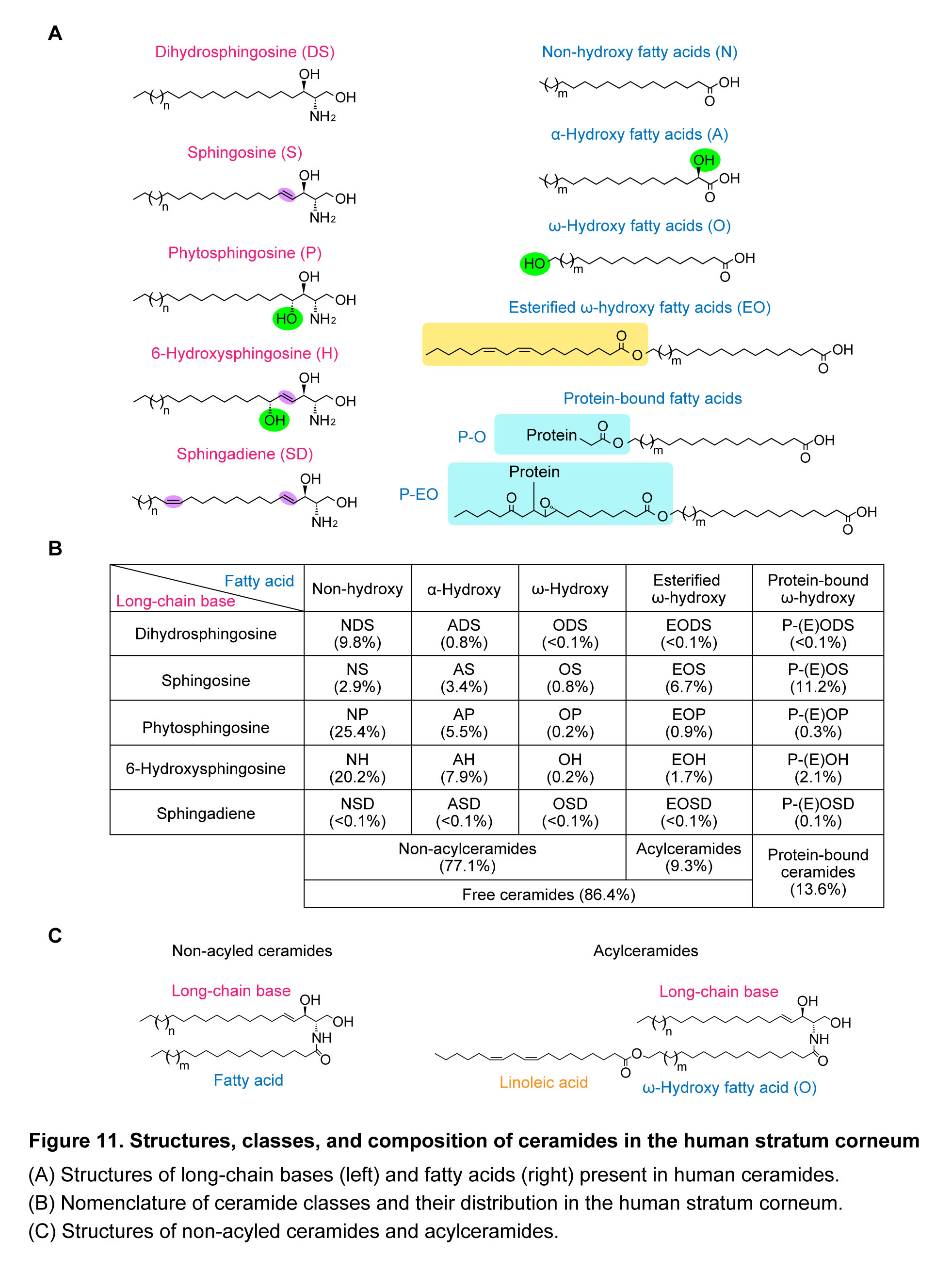
Ceramides consist of a long-chain base and a fatty acid linked via an amide bond. In humans, 25 classes of ceramides are formed by combinations of five types of long-chain bases (sphingosine [S], dihydrosphingosine [DS], phytosphingosine [P], 6-hydroxysphingosine [H], and 4,14-sphingadiene [SD]) and five types of fatty acids (non-hydroxy fatty acids [N], α-hydroxy fatty acids [A], ω-hydroxy fatty acids [O], esterified ω-hydroxy fatty acids [EO], and protein-bound fatty acids [PB- (P-O/P-EO)]). Each ceramide class is designated by the combination of abbreviations for the fatty acid and long-chain base (e.g., NS and EOS; Figure 11A, B). Ceramides containing EO-type fatty acids are referred to as acylceramides (more precisely, ω-O-acylceramides), and the esterified fatty acid is predominantly linoleic acid (Figure 11C). Among free (non-protein-bound) ceramides, those other than acylceramides are collectively referred to as non-acylated ceramides, which correspond to what are generally referred to as ceramides. Many of the ceramides present in the lipid lamellae are non-acylated ceramides. Although acylceramides are present in smaller amounts than non-acylated ceramides, their unique structural properties are critical for the formation and maintenance of the multilamellar organization of the lipid lamellae, and thus for skin barrier formation. A fraction of acylceramides is converted into protein-bound ceramides. During this process, the linoleate moiety undergoes sequential modifications: hydroperoxidation, isomerization, and oxidation, ultimately yielding epoxy–enone-containing acylceramides. Regarding the structure of protein-bound ceramides, a long-standing model (Figure 11A; the protein-bound ω-hydroxy fatty acid [P-O] model) proposes that, following the removal of the modified linoleate moiety by hydrolysis, the resulting ω-hydroxy ceramides become covalently linked via their ω-hydroxyl groups to glutamate residues of corneocyte surface proteins (cornified envelope proteins) through ester bond formation. In contrast, a model proposed in 2020 (Figure 11A; the protein-bound modified linoleate-esterified ω-hydroxy fatty acid [P-EO] model) suggests that acylceramides containing an epoxy–enone-modified linoleate moiety undergo covalent attachment to proteins through reaction of the enone group, such as Michael addition or Schiff base formation. We subsequently demonstrated, using mouse epidermis, the presence of P-EO-type protein-bound ceramides and showed that they are of the epoxy-ketone type, in which the modified linoleate moiety is linked to cysteine residues via Michael addition1). In contrast, under our experimental conditions, neither the conventionally proposed P-O-type protein-bound ceramides nor P-EO-type ceramides linked to amino acid residues other than cysteine were detected. We then analyzed the structures of protein-bound ceramides in the human stratum corneum and found that the epoxy-ketone type, which is the predominant form in mouse epidermis, is present only at low levels in humans. Instead, dihydroxy-ketone-type P-EO ceramides, in which the epoxy group has been hydrolyzed, were found to constitute the major form of protein-bound ceramides in the human stratum corneum (Figure 11A)2).
In most previous analytical approaches, P-O-type and P-EO-type protein-bound ceramides have not been distinguished and have instead been collectively measured as protein-bound ceramides. In such cases, these species are collectively denoted as PB- (as in Figure 4) or as P-(E)O (as in Figure 11B). In these methods, the fraction containing protein-bound ceramides, obtained after removing free ceramides from the epidermis or stratum corneum, is subjected to alkaline treatment. This procedure releases all protein-bound ceramides as ω-hydroxy ceramides (O-type ceramides), which are then measured. In contrast, selective measurement of P-EO-type protein-bound ceramides can be achieved by releasing them from the protein-bound ceramide fraction via oxidative sulfoxide elimination as epoxy-enone acylceramides (for epoxy-ketone-type protein-bound ceramides) or dihydroxy-enone acylceramides (for dihydroxy-ketone-type protein-bound ceramides) prior to analysis1, 2).
Ceramide composition in mammalian tissues and the stratum corneum
In most mammalian tissues, ceramide composition is relatively simple, and only a limited number of ceramide classes—such as NS, NDS, NSD, and AS—are present. NS is the predominant ceramide class across most tissues. NSD is also widely distributed but present at lower levels than NS, while it shows relatively high abundance in the kidney and, to a lesser extent, in the brain3). NP is preferentially found in epithelial tissues, including the epidermis, esophagus, stomach, small intestine, large intestine, and kidney4). AS is enriched in brain myelin and is also found in the epidermis, stomach, and submandibular glands5). In contrast, the epidermis, especially the stratum corneum, contains a large diversity and abundance of ceramides. Acylceramides, protein-bound ceramides, and H-type ceramides containing 6-hydroxysphingosine (H) are unique to the epidermis (although acylceramides and protein-bound ceramides are also present in stratified squamous epithelia such as the oral cavity and esophagus; see below). Each ceramide class comprises multiple species that differ in the chain lengths of both fatty acids and long-chain bases, as well as the degree of unsaturation of fatty acids. We established a mass spectrometry–based method (liquid chromatography–tandem mass spectrometry [LC–MS/MS]; for details, see the section “Analysis of lipids by LC-MS/MS” below) that enables the separation and comprehensive quantification of ceramide classes and species in the stratum corneum. Using this approach, we first showed in 2020 that, when the long-chain base was limited to C18 species, the human stratum corneum contains 26 classes/408 species of ceramides, whereas mouse epidermis contains 21 classes/342 species6). Subsequently, in 2022, by expanding the analysis to include long-chain bases ranging from C16 to C26, we identified 23 classes/1,581 species of ceramides in the human stratum corneum, thereby elucidating the overall ceramide composition of this tissue7). The apparent decrease in the number of human ceramide classes in the latter analysis is due to the exclusion of minor classes. In this analysis, protein-bound ceramides were converted to O-type ceramides by alkaline treatment prior to measurement, as described above.
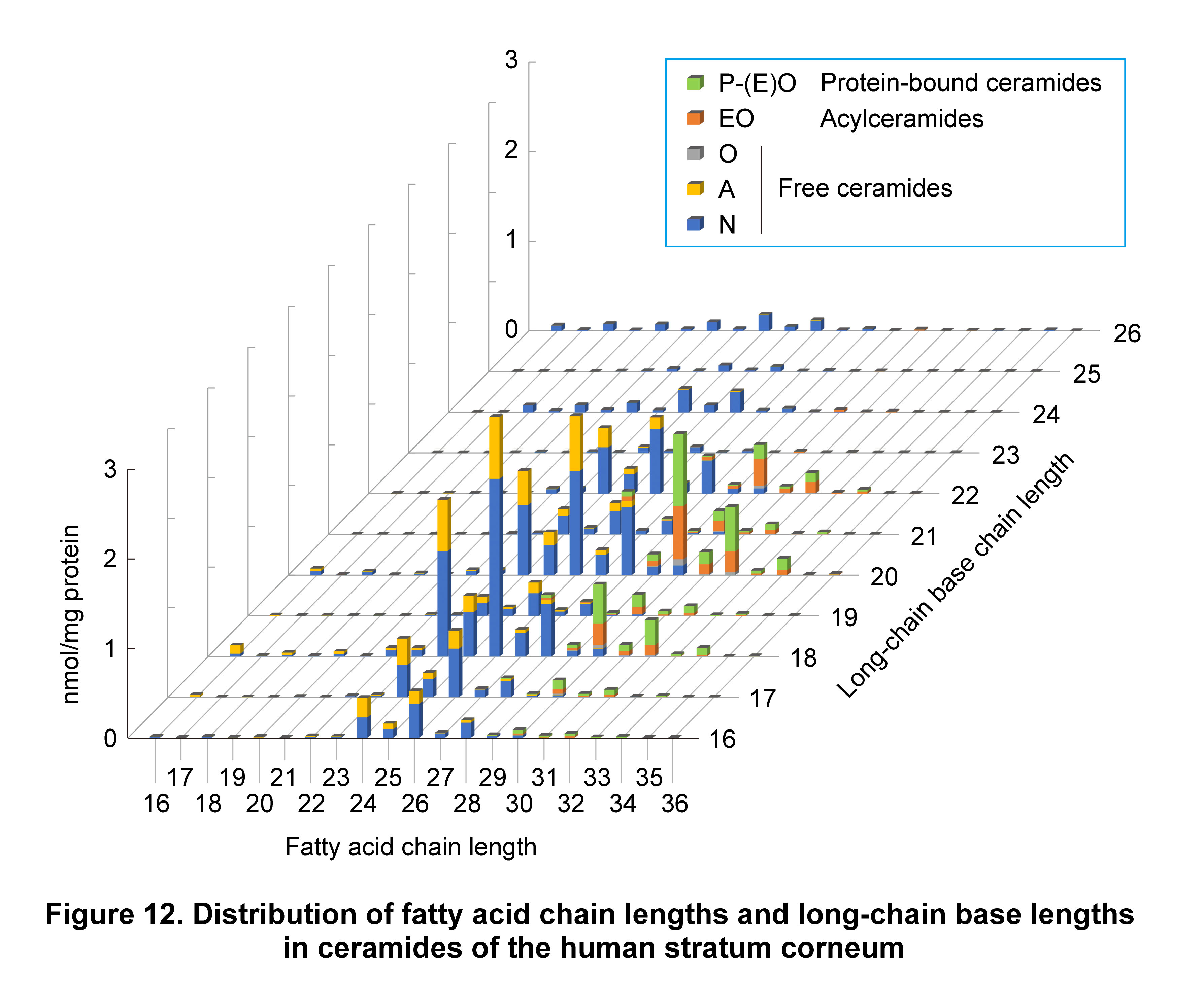
The fatty acid chain lengths of ceramides in most tissues generally range from C16 to C24, whereas the long-chain bases are almost exclusively limited to C18. In contrast, ceramides in the human stratum corneum exhibit a much wider range, with fatty acid chain lengths ranging from C16 to C36 and long-chain bases from C16 to C26 (Figure 12). The chain length distribution of fatty acids in stratum corneum ceramides varies depending on the ceramide class. In non-acylated ceramides, N-type and A-type ceramides, which contain non-hydroxy fatty acids and α-hydroxy fatty acids, respectively, show fatty acid chain lengths ranging from C16 to C28, with C24 and C26 being predominant. In contrast, the fatty acid moieties (ω-hydroxy fatty acids) in O-type ceramides, acylceramides, and protein-bound ceramides are longer, ranging from C28 to C36, with C30, C32, and C34 being predominant. The difference in fatty acid chain lengths between ceramides in general tissues and those in the stratum corneum likely reflects the difference in thickness between the lipid bilayer of cellular membranes (~10 nm) and the long periodicity phase (~13 nm) of the lipid lamellae in the stratum corneum. The presence of diverse ceramide species in the human stratum corneum may contribute to adaptation to environmental changes. The lipid lamellae are in a gel phase and must remain in this state under varying environmental conditions, including changes in temperature, pH, humidity, and ionic strength. Heterogeneity in lipid composition broadens the phase transition temperature range, thereby allowing the gel phase of the lipid lamellae to be maintained over a wide range of conditions. The high cholesterol content in the stratum corneum may also facilitate gel phase formation through its strong affinity for ceramides.
A characteristic feature of human stratum corneum ceramides is the abundance of P-type and H-type ceramides, which contain phytosphingosine (P) or 6-hydroxysphingosine (H), respectively. Their relative proportions of total ceramides are 25.4% for NP, 20.2% for NH, 5.5% for AP, and 7.9% for AH, together accounting for approximately 60% (Figure 11B)7). These ceramides contain one (NP and NH) or two (AP and AH) additional hydroxyl groups compared with NS, which is predominant in most tissues. Hydrogen bonding mediated by these hydroxyl groups is likely to enhance lipid–lipid interactions within the lipid lamellae, and the increase in hydroxyl groups may also confer the polarity required for the stabilization of the lamellar structure in the lipid lamellae. In contrast, SD-type ceramides, which contain 4,14-sphingadiene (SD), the only long-chain base with a cis double bond, are present at low levels in the stratum corneum. This is likely because the cis double bond at the C-14 position interferes with tight lipid packing within the lipid lamellae. In patients with atopic dermatitis, decreases in total ceramide levels, alterations in ceramide class composition, and shortening of fatty acid chain lengths have been reported. Among these, changes in class composition include reductions in non-acylated ceramides with an additional hydroxyl group in the long-chain base (NP and NH) as well as in acylceramides (EOS, EOH, and EOP). Similar changes in ceramide class composition are observed not only in pathological conditions such as atopic dermatitis but also, to a lesser extent, in nonpathological conditions such as dry skin (for details, see below)8).
Degradation of ceramides in keratinocytes
We have demonstrated that ceramides in keratinocytes are not only synthesized but also degraded9). This degradation is mediated primarily by ASAH1, an acid ceramidase. ASAH1 exhibits distinct substrate specificities toward different ceramide classes; it shows high activity toward NS and NDS, weaker activity toward NP and NH, and little to no activity toward AP and AH9). In other words, ASAH1 preferentially acts on ceramide classes with fewer hydroxyl groups and shows lower activity toward those with more hydroxyl groups. As described above, ceramides containing additional hydroxyl groups are enriched in the stratum corneum and contribute to strong lipid–lipid interactions within the lipid lamellae and increased polarity of ceramides. Taken together, our results indicate that both ceramide synthesis and class-dependent degradation by ASAH1 are involved in maintaining the appropriate ceramide class composition in the stratum corneum.
Ceramide hydroxylation/desaturation enzymes DEGS1 and DEGS2
S-type ceramides containing sphingosine (S) and P-type ceramides (phytoceramides) containing phytosphingosine (P) are generated from DS-type ceramides (dihydroceramides), which contain dihydrosphingosine (DS), through C-4 desaturation or C-4 hydroxylation, respectively. The enzymes responsible for these reactions are DEGS1 and DEGS2. DEGS1 functions exclusively as a C-4 desaturase, whereas DEGS2 is a bifunctional enzyme exhibiting both C-4 desaturase and C-4 hydroxylase activities. We found that DEGS2 exhibits chain-length-dependent reaction preferences for DS-type ceramide substrates; hydroxylation predominates for those with very-long-chain fatty acids, whereas desaturation is favored for those with shorter fatty acids4). As described above, P-type ceramides generated by DEGS2, such as NP and AP, represent the most abundant ceramide classes in the human stratum corneum and likely play important roles in skin barrier function. In contrast, P-type ceramide levels in the mouse epidermal stratum corneum are low (approximately 1% of total ceramides), and Degs2 KO mice do not exhibit skin barrier defects4). This observation suggests that the low abundance of P-type ceramides renders them dispensable for skin barrier function in mice.
Acylceramide biosynthetic pathway and genes
Acylceramides are involved in the formation and maintenance of the lipid lamellae, whereas protein-bound ceramides contribute to the formation of the CLE; therefore, both are essential for skin barrier formation. Most genes involved in acylceramide and protein-bound ceramide synthesis have been identified since 2010. Mutations in these genes cause congenital ichthyosis in humans, and knockout (KO) mice for these genes exhibit neonatal lethality due to severe skin barrier defects. We have identified five of the seven genes involved in acylceramide biosynthesis (ELOVL1, CYP4F22, FATP4, PNPLA1, and ABHD5) and have elucidated the details of the biosynthetic pathway, including the substrates, reaction order, and reaction types. The reactions in this pathway are shown below and in Figure 13.
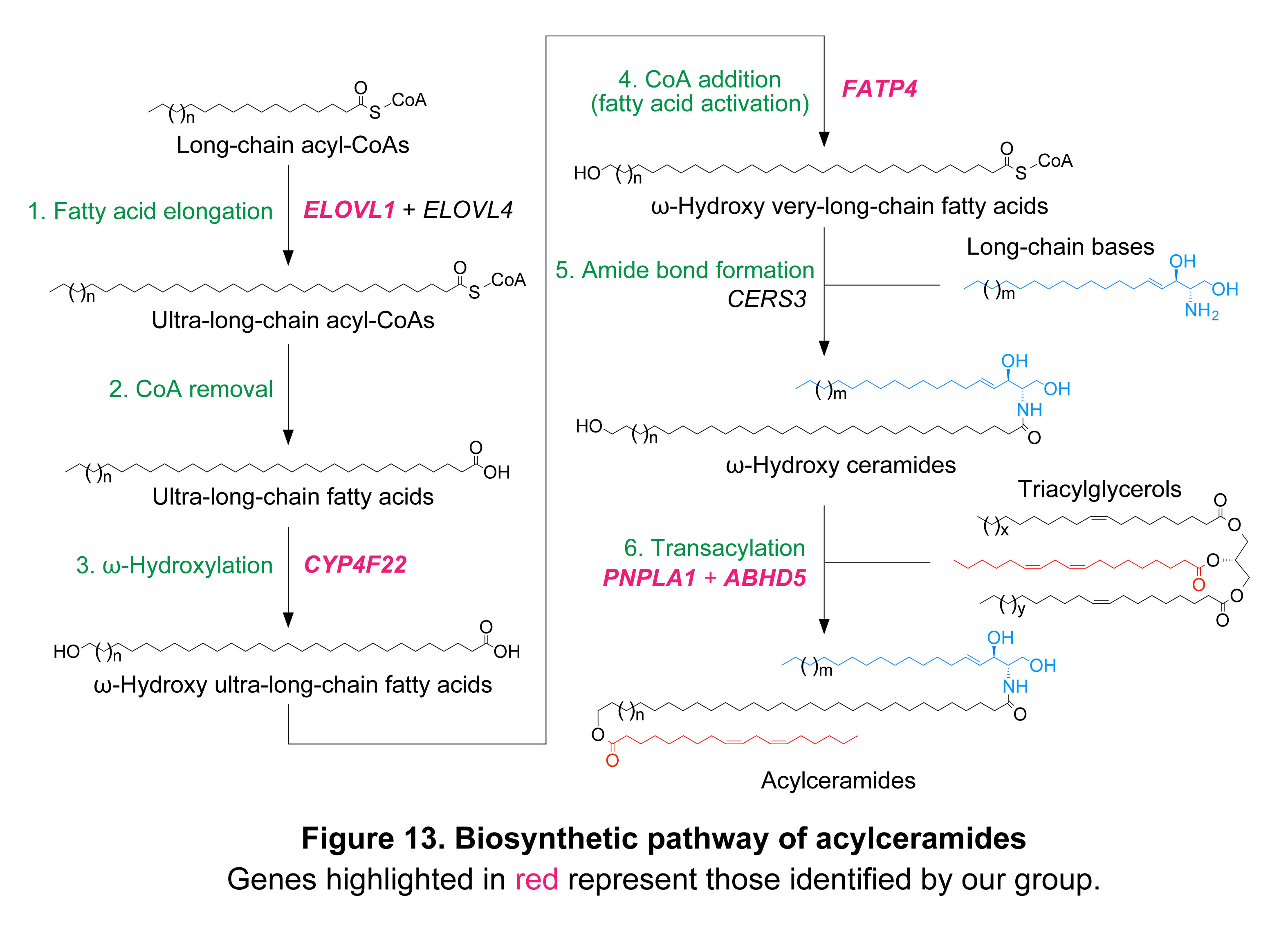
- The fatty acid elongases ELOVL1 and ELOVL4 catalyze the elongation of acyl-CoAs up to C26 and C30–36, respectively. We demonstrated that ELOVL1 exhibits activity toward C20–26 acyl-CoAs in vitro, producing C22–28 acyl-CoAs10). In these reactions, ELOVL1 shows higher activity toward C22:0-CoA and C24:0-CoA, generating C24:0-CoA and C26:0-CoA, respectively, compared with other ELOVLs. We next generated Elovl1 KO mice and showed that they exhibit neonatal lethality associated with skin barrier defects11). In the epidermis of these mice, a marked reduction in ≥C26 ceramides (including acylceramides), a moderate decrease in C24 ceramides, and an increase in ≤C22 ceramides were observed12). These findings indicate that ELOVL1 plays a particularly important role in the elongation of C22-CoA to C26-CoA in the epidermis. In addition, we reported for the first time that mutations in ELOVL1 in humans cause a neurocutaneous disorder associated with ichthyosis (IKSHD syndrome; ichthyotic keratoderma, spasticity, hypomyelination, and dysmorphia)13), and we showed that acylceramide levels are greatly reduced in the stratum corneum of patients with IKSHD syndrome14).
- Production of ultra-long-chain fatty acids from ultra-long-chain acyl-CoAs via hydrolysis: the thioesterase responsible for this reaction remains to be identified, although it is possible that members of the ACOT (acyl-CoA thioesterase) family proteins, which comprise approximately 10 members in humans, function redundantly.
- Production of ω-hydroxy ultra-long-chain fatty acids via ω-hydroxylation of ultra-long-chain fatty acids: the cytochrome P450 enzyme CYP4F22 (CYP4F39 in mice) catalyzes the ω-hydroxylation of ultra-long-chain fatty acids. Although CYP4F22 had been known as a causative gene of autosomal recessive congenital ichthyosis (ARCI), the mechanism by which mutations in CYP4F22 lead to ichthyosis remained unclear. We demonstrated that CYP4F22 catalyzes this reaction through analyses using a cell-based expression system, an in vitro assay, activity measurements of ichthyosis-associated mutants, and analysis of acylceramide levels in the stratum corneum of patients with CYP4F22 mutations15–17). We next generated Cyp4f39 KO mice and showed that they exhibit neonatal lethality associated with skin barrier defects, primarily due to impaired acylceramide production18). Together, these analyses reveal that defective acylceramide production is a major cause of the pathogenesis of ichthyosis.
- Production of ω-hydroxy ultra-long-chain acyl-CoAs via CoA addition to ω-hydroxy ultra-long-chain fatty acids: this reaction is catalyzed by the acyl-CoA synthetase FATP4 (also known as SLC27A4 and ACSVL4). Although FATP4 had been identified as the causative gene of ichthyosis prematurity syndrome, the molecular mechanism by which its mutations cause the disease remained unclear. We demonstrated that FATP4 functions as an acyl-CoA synthetase that converts ω-hydroxy ultra-long-chain fatty acids into their acyl-CoA forms in the acylceramide biosynthetic pathway, based on in vitro analysis and the observation of a marked reduction in acylceramide levels in Fatp4 KO mice we generated19).
- Production of ω-hydroxy ceramides via condensation of ω-hydroxy ultra-long-chain acyl-CoAs with long-chain bases: this reaction is catalyzed by the ceramide synthase CERS3. We were the first to report the enzymatic activity of CERS3 as a ceramide synthase20). In this study, we found that CERS3 exhibits activity toward acyl-CoAs with a wide range of chain lengths from C16 to C26 in in vitro assays. Activities toward substrates with chain lengths of ≥C28 could not be analyzed due to the unavailability of the corresponding acyl-CoAs. We then confirmed, using a cell-based assay with HEK293T cells, that CERS3 is involved in the production of ceramides with a broad range of fatty acid chain lengths21). Furthermore, we found that CERS3 expression increases during keratinocyte differentiation and that its expression levels are markedly higher than those of other CERS genes in differentiated human keratinocytes22). These findings suggest that CERS3 has low substrate specificity for acyl-CoA chain length, enabling the synthesis of ceramides with a wide range of chain lengths and potentially contributing to the production of a broad spectrum of epidermal ceramides, including acylceramides. The involvement of CERS3 in acylceramide production was subsequently demonstrated by other researchers through analyses of Cers3 KO mice. CERS3 is a causative gene of ARCI in humans, and we profiled ceramides in the stratum corneum of patients with CERS3 mutations and found that acylceramide levels are reduced23).
- Production of acylceramides via ester bond formation between ω-hydroxy ceramides and linoleic acid: this reaction is catalyzed by the transacylase PNPLA1, and ABHD5 enhances substrate utilization by PNPLA1. PNPLA1 and ABHD5 had been known as causative genes of ARCI and Chanarin–Dorfman syndrome, an ichthyosis-associated lipid storage disorder, respectively; however, the molecular mechanisms by which their mutations lead to these ichthyoses remained unclear. We demonstrated that PNPLA1 functions as an enzyme catalyzing the final step of acylceramide production through cell-based assays (overexpression and knockdown), in vitro analyses, and functional analyses of ichthyosis-associated mutants24, 25). In particular, in the in vitro experiment, we showed that PNPLA1 utilizes triacylglycerols containing linoleic acid rather than linoleoyl-CoA as substrates, indicating that it acts as a transacylase rather than an acyltransferase24). We also revealed that ABHD5 enhances PNPLA1-catalyzed acylceramide production by facilitating the utilization of triacylglycerols as substrates26). In cell-based assays, acylceramide production by PNPLA1 was enhanced by ABHD5 expression, whereas ABHD5 missense mutants associated with Chanarin–Dorfman syndrome did not exhibit this stimulatory effect26). PNPLA1 alone showed a dispersed localization pattern throughout the cell, whereas co-expression with ABHD5 recruited PNPLA1 to lipid droplets, where its substrate triacylglycerols are stored26). We also demonstrated that acylceramide levels are reduced in the stratum corneum of patients with PNPLA1 mutations27).
Phenotypes of KO mice for acylceramide biosynthetic genes
We generated and analyzed Elovl1, Cyp4f39, and Fatp4 KO mice, all of which exhibited neonatal lethality with severe skin barrier defects11, 18, 19). Acylceramide levels were markedly reduced in these KO mice, to 0.6%, 1.6%, and 12% of wild-type levels in Cyp4f39 KO, Elovl1 KO, and Fatp4 KO mice, respectively12). Because protein-bound ceramides are derived from acylceramides, their levels were also decreased in these KO mice, to 0.2%, 30%, and 33%, respectively. The severity of skin barrier defects in these mice was in the order of Cyp4f39 KO, Elovl1 KO, and Fatp4 KO mice. This difference can largely be explained by the extent of reduction in acylceramide and protein-bound ceramide levels, but is also partly attributable to KO gene-dependent alterations in the composition of free ceramides other than acylceramides. Because KO mice for all genes involved in acylceramide and protein-bound ceramide production reported to date, including those generated by other researchers as well as our own, exhibit neonatal lethality, the importance of acylceramides and protein-bound ceramides in maintaining the skin barrier in adult mice remains unclear. To address this issue, we generated and analyzed tamoxifen-inducible conditional KO (cKO) mice, in which Elovl1 can be deleted in adult mice28). In Elovl1 cKO mice, acylceramide levels began to decrease from day 5 after tamoxifen administration and were reduced to approximately 50% of control levels on day 10. At this time point, impaired formation of lipid lamellae in the stratum corneum and epidermal thickening were observed. By day 15, acylceramide and protein-bound ceramide levels were further reduced to approximately 30% of control levels, and transepidermal water loss was increased, indicating impaired skin barrier function. These findings indicate that both acylceramides and protein-bound ceramides play important roles in maintaining the skin barrier in adult mice. However, the lethality observed in neonatal KO mice for acylceramide biosynthetic genes was not observed even after prolonged tamoxifen administration in adult Elovl1 cKO mice. This difference may reflect the increasing contribution of hair and sebum to the permeability barrier during postnatal development in hair-covered animals such as mice, which may reduce the relative importance of the stratum corneum in the skin barrier.
NIPAL4 mutation–induced pathogenesis of ichthyosis
Mutations in genes involved in the synthesis of acylceramides or protein-bound ceramides cause congenital ichthyosis in humans. However, among causative genes for ichthyosis, some remained poorly characterized, and the mechanisms by which their mutations cause ichthyosis remained unclear. Such ichthyosis-causative genes include NIPAL4 and ALDH3A2, responsible for ARCI and Sjögren–Larsson (SLS), an ichthyosis-associated syndrome, respectively. To elucidate the mechanisms underlying ichthyosis associated with mutations in these genes, we analyzed Nipal4 KO mice and SLS model mice. Nipal4 KO mice showed reduced levels of acylceramide (ω-O-acylceramide) in the epidermis29). In addition, the composition of the O-acyl chain was altered. In wild-type mice, the major O-acyl chain of ω-O-acylceramides was linoleic acid (89%), whereas this proportion was reduced to 66% in KO mice, accompanied by an increase in other fatty acids, particularly oleic acid, which increased from 2% to 16%30). Moreover, the production of another class of acylceramides, 1-O-acylceramides, was induced in the epidermis of Nipal4 KO mice. NIPAL4 encodes a Mg2+ transporter. In keratinocytes derived from wild-type mice, intracellular Mg2+ levels increased during differentiation, whereas such an increase was not observed in Nipal4 KO keratinocytes29). Mg2+ is known to stabilize membranes through interactions with phospholipids. In keratinocytes of the stratum granulosum, which produce specialized lipids such as ω-O-acylceramides containing ultra-long-chain acyl moieties, this increase in intracellular Mg2+ is thought to help stabilize intracellular membranes, particularly the endoplasmic reticulum, where these lipids are synthesized. In Nipal4 KO mice, the normal increase in intracellular Mg2+ does not occur, resulting in altered membrane properties compared with those in wild-type stratum granulosum keratinocytes, which in turn may affect lipid metabolism and lead to the production of abnormal lipid species.
Pathogenesis of ichthyosis in Sjögren–Larsson syndrome (SLS)
ALDH3A2 encodes fatty aldehyde dehydrogenase. We have demonstrated that ALDH3A2 is involved in the metabolism of long-chain aldehydes generated during the degradation of long-chain bases (for details, see Section 2. Physiological functions and metabolism of the bioactive lipid sphingosine-1-phosphate). We generated Aldh3a2 KO mice; however, unlike SLS patients, these mice did not exhibit skin barrier defects under normal conditions31). Nevertheless, when the stratum corneum was perturbed, Aldh3a2 KO mice showed delayed recovery of the skin barrier compared with wild-type mice31). The absence of skin barrier defects under normal conditions is likely because mice possess Aldh3b2, which has a function redundant with Aldh3a232). In contrast, ALDH3B2 is a pseudogene in humans. We further generated Aldh3a2 Aldh3b2 double KO mice and found that these mice exhibited an ichthyosis-like phenotype accompanied by reduced levels of acylceramide in the epidermis33). We propose that accumulated long-chain aldehydes impair enzymes involved in acylceramide production, leading to decreased acylceramide levels. Furthermore, reduced acylceramide levels were also observed in ALDH3A2-deficient human keratinocytes and in patients with SLS33, 34). Together, our findings indicate that, similar to the case of ARCI caused by NIPAL4 mutations described above, ichthyosis in SLS caused by ALDH3A2 mutations is also due to impaired acylceramide production. Thus, although many ichthyosis-causative genes directly impair acylceramide synthesis, mutations in other genes can lead to ichthyosis by indirectly affecting acylceramide production.
Profiling of stratum corneum ceramides in congenital ichthyosis and dry skin
We have previously performed ceramide profiling of the stratum corneum in several patients with congenital ichthyosis as well as individuals with dry skin. The ceramide composition in patients with ichthyosis differed depending on the mutated gene, with an increase in the substrates of the reactions catalyzed by the gene products and a decrease in the corresponding products and their metabolites. In contrast, dry skin exhibited changes in free ceramide composition similar to those observed in atopic dermatitis, although to a lesser extent, namely decreases in NP, EOS, and EOP, and increases in NS and AS8). Although protein-bound ceramide composition in atopic dermatitis has not been examined, we found a decrease in PB-H and an increase in PB-SD in dry skin. The congenital ichthyosis patients we have profiled carried mutations in the following genes: ALDH3A2 (SLS)34), CERS3 (ARCI)23), ELOVL1 (IKSHD syndrome)14), CYP4F22 (ARCI)15, 17), PNPLA1 (ARCI)27), and SDR9C7 (ARCI)35). Among these, CERS3, ELOVL1, CYP4F22, and PNPLA1 are directly involved in acylceramide synthesis as described above, and patients with mutations in these genes exhibited reduced levels of acylceramides14, 15, 17, 23, 27, 34). In contrast, SDR9C7, which encodes a short-chain dehydrogenase/reductase family protein, is involved in the formation of protein-bound ceramides, and mutations in this gene resulted in decreased levels of protein-bound ceramides35).
Oral barrier formation by ceramides
Acylceramides and protein-bound ceramides were previously thought to be present only in the epidermis; however, we found that these lipids are also present in the stratified epithelial tissues from the oral cavity to the forestomach in mice, where they play important roles in the formation of the oral barrier36). In cKO mice lacking the fatty acid elongase Elovl1 in these tissues, morphological changes in the esophagus and tongue, including hyperkeratosis and alterations in epithelial structure, as well as increased permeability to fluorescent dyes and avoidance behavior toward capsaicin-containing water, were observed. Furthermore, in humans, acylceramides were detected at least in the buccal mucosa and gingiva we analyzed, and protein-bound ceramides were detected in the gingiva. These findings indicate that protein-bound ceramides are present in stratified keratinized squamous epithelia. Thus, the importance of ceramides in permeability barrier formation, which had been considered to be limited to the epidermis, extends to the oral cavity (and likely the upper digestive tract).
Analysis of lipids by LC–MS/MS
Lipids containing ceramides have been analyzed using various methods, including thin-layer chromatography, mass spectrometry (MS), nuclear magnetic resonance (NMR), and infrared spectroscopy. Among these, MS and tandem mass spectrometry (MS/MS) represent the gold standard for analysis. In MS, molecules are separated according to mass (more precisely, the mass-to-charge ratio; m/z), whereas in MS/MS, selected precursor ions are fragmented by collision with an inert gas to generate product ions, which are then detected. For lipid ionization, electrospray ionization (ESI) is commonly used, whereas atmospheric pressure chemical ionization (APCI) is used for less polar lipids. MS (/MS) is performed either by direct sample introduction (shotgun method) or after separation by liquid chromatography (LC). Untargeted lipidomics using the shotgun method offers high coverage but is susceptible to ion suppression, which may compromise quantification accuracy and specificity. Separation of molecules by LC prior to ionization is effective in reducing ion suppression caused by contaminants and improving the quantitative accuracy and sensitivity of detected molecules. After LC separation, both targeted lipidomics (enabling highly sensitive and specific quantification of individual lipid species) and untargeted lipidomics (comprehensive lipid detection) can be performed. LC using normal-phase columns is effective for separating lipid classes with different polarities, whereas reversed-phase columns are suitable for separating lipid species with different carbon chain lengths (and thus hydrophobicity).
In reversed-phase LC-MS, separation is achieved based on differences in mass and hydrophobicity; however, many ceramide species share identical mass and hydrophobicity and therefore cannot be distinguished by this method. For example, ceramides with a total carbon number of C42 and an unsaturation of 2 (C44:2; [M+H]+ = 648) include an NS species composed of d18:1 sphingosine and a C24:1 fatty acid (d18:1/C24:1), which cannot be distinguished from other NS species with the same total carbon number and unsaturation (e.g., d20:1/C22:1 and d22:1/C20:1). Therefore, LC-MS/MS is essential for distinguishing these species. MS/MS includes several measurement modes, such as precursor ion scanning, neutral loss scanning, product ion scanning, and multiple reaction monitoring (MRM). Among these, MRM provides high specificity and sensitivity by selectively detecting predefined precursor–product ion m/z pairs (set in Q1 and Q3, respectively). By appropriately selecting Q3 in MRM, individual species within C44:2 ceramides can be distinguished and quantified (e.g., d18:1/C24:1: Q3 = 264.3; d20:1/C22:1: 292.3; d22:1/C20:1: 320.3).
In the analysis of ceramides, attention should be paid to fragmentation during ionization (in-source fragmentation). Ceramides, particularly those containing sphingosine or 6-hydroxysphingosine, readily undergo dehydration6). Accordingly, depending on the ceramide class, it is necessary to appropriately select the Q1 target as either the intact ion [M+H]+ or the dehydrated ion [M+H−H2O]+.
Product ions of ceramides differ depending on the long-chain base moiety. Taking advantage of this characteristic, setting the Q3 value to an m/z characteristic of each long-chain base is effective (for C18 long-chain bases: NS, 264.3; NDS, 284.3; NP, 300.3; NH, 280.3; NSD, 262.3)6).
Another important point regarding in-source fragmentation is that glycosphingolipids, such as glucosylceramides, can be converted into ceramides during ionization. This conversion is particularly prominent for glucosyl NP ceramides, many of which are detected as ceramides. However, ceramides and glucosylceramides can be distinguished based on their clearly different retention times in LC.
As described above, many ceramide species can share the same Q1 value. In the case of [M+H]+ = 648, not only are multiple species present within C44:2 NS, but other classes are also possible, such as the dehydrated forms of d18:1/C24:0 AS, d18:0/C25:0 NDS, d18:0/C24:1 ADS, t18:0/C24:1 NP, t18:1/C24:0 NH, and t18:1/C23:1 AH, as well as the corresponding deglycosylated forms of glucosylceramides, at least theoretically (including in-source fragmentation events that are unlikely to occur in practice). Therefore, to distinguish ceramide species, retention time in LC together with appropriate Q1 and Q3 settings is essential. In addition, optimizing collision energy for each species is important for accurate quantification. Even within the same ceramide class, species with longer fatty acyl chains require higher collision energy. Once these parameters are appropriately set, comprehensive and highly sensitive quantification of ceramide species becomes possible. Indeed, using LC-MS/MS in MRM mode with a triple quadrupole mass spectrometer and a reversed-phase column, we established a highly accurate method for ceramide analysis and successfully quantified more than 1,500 ceramide species in human stratum corneum7).
Another important aspect in LC-MS or LC-MS/MS analysis is ensuring the reliability of peak assignment. Even in highly specific MRM measurements, peaks are not necessarily singular, and multiple peaks are often detected. To accurately assign peaks corresponding to the target lipid species, preliminary studies using chemically synthesized standards, including the determination of retention time, characterization of product ions, and optimization of MS conditions, are critically important. We use deuterium-labeled ceramides as internal standards for the measurement of a wide range of ceramide classes. However, for each class, the available internal standards are often single species with specific chain lengths, and the retention times of other species in LC must therefore be estimated based on their chain lengths and positions of double bonds. For example, under our LC conditions, an increase of two carbons in chain length results in an approximately 0.3 min increase in retention time, whereas introduction of a single cis double bond at the ω9 position shortens the retention time by approximately 0.3 min. In addition, for peaks with insufficient assignment reliability, product ion scanning should be performed to confirm the identity of the target molecule based on its fragmentation pattern. Biological approaches can also aid in validating peak assignment. For example, when the target lipid is expected to decrease or disappear in KO mice for a specific gene, comparison with wild-type mice can be used to confirm the assignment. From a quantitative perspective, the use of lipid standards not present in biological samples, such as deuterium-labeled ceramides, is useful for accurate quantification.
While we take these analytical considerations into account in our ceramide measurements, in a considerable number of reports by other researchers, peak assignments appear to be incorrect, leading to the measurement of lipids other than ceramides. In addition, even when peaks are correctly assigned to the intended ceramide species, the reliability of quantification is often compromised by factors such as ion suppression, in-source fragmentation, and inappropriate collision energy settings. Therefore, when referring to published studies, careful evaluation of both the measurement methods and the validity of peak assignment is important.
Conclusion
In summary, we have elucidated the diversity of stratum corneum ceramides, identified numerous ceramide-related genes (for details, see Section 1. Identification of sphingolipid-related genes), and generated and analyzed genetically modified mouse models of these genes, as well as performed ceramide profiling in the stratum corneum of patients with ichthyosis and individuals with dry skin. Through these studies, we have clarified the molecular mechanisms underlying the generation of ceramide diversity, its physiological significance, and its involvement in disease pathology. Based on these findings and the techniques we have established, we aim to develop novel diagnostic and therapeutic approaches for skin diseases, including atopic dermatitis and ichthyosis.
References
- Ohno Y, Nakamura T, Iwasaki T, Katsuyama A, Ichikawa S, Kihara A. (2023) Determining the structure of protein-bound ceramides, essential lipids for skin barrier function. iScience, 26, 108248.
- Kojima A, Sugiyama T, Ito Y, Otsuka K, Ohno Y, Kihara A. (2026) Discovery of dihydroxy-ketone–type protein-bound ceramides as the dominant type in human stratum corneum. FASEB J, 40, e72184.
- Jojima K, Edagawa M, Sawai M, Ohno Y, Kihara A. (2020) Biosynthesis of the anti-lipid-microdomain sphingoid base 4,14-sphingadiene by the ceramide desaturase FADS3. FASEB J, 34, 3318–3335.
- Ota A, Morita H, Naganuma T, Miyamoto M, Jojima K, Nojiri K, Matsuda J, Kihara A. (2023) Bifunctional DEGS2 has higher hydroxylase activity toward substrates with very-long-chain fatty acids in the production of phytosphingosine ceramides. J Biol Chem, 299, 104603.
- Mori K, Naganuma T, Kihara A. (2023) Role of 2-hydroxy acyl-CoA lyase HACL2 in odd-chain fatty acid production via α-oxidation in vivo. Mol Biol Cell, 34, ar85.
- Kawana M, Miyamoto M, Ohno Y, Kihara A. (2020) Comparative profiling and comprehensive quantification of stratum corneum ceramides in humans and mice by LC/MS/MS. J Lipid Res, 61, 884–895.
- Suzuki M, Ohno Y, Kihara A. (2022) Whole picture of human stratum corneum ceramides, including the chain-length diversity of long-chain bases. J Lipid Res, 63, 100235.
- Akiyama F, Takahashi N, Ueda Y, Tada S, Takeuchi N, Ohno Y, Kihara A. (2024) Correlations between skin condition parameters and ceramide profiles in the stratum corneum of healthy individuals. Int J Mol Sci, 25, 8291.
- Nobumoto W, Naganuma T, Nozaka N, Ohno Y, Nojiri K, Kihara A. (2026) Acid ceramidase ASAH1 is a key regulator of epidermal ceramide levels and composition. J Biol Chem, 302, 111178.
- Ohno Y, Suto S, Yamanaka M, Mizutani Y, Mitsutake S, Igarashi Y, Sassa T, Kihara A. (2010) ELOVL1 production of C24 acyl-CoAs is linked to C24 sphingolipid synthesis. Proc Natl Acad Sci USA, 107, 18439–18444.
- Sassa T, Ohno Y, Suzuki S, Nomura T, Nishioka C, Kashiwagi T, Hirayama T, Akiyama M, Taguchi R, Shimizu H, Itohara S, Kihara A. (2013) Impaired epidermal permeability barrier in mice lacking Elovl1, the gene responsible for very-long-chain fatty acid production. Mol Cell Biol, 33, 2787–2796.
- Yamamoto Y, Sassa T, Kihara A. (2024) Comparison of skin barrier abnormalities and epidermal ceramide profiles among three ω-O-acylceramide synthesis-deficient mouse strains. J Dermatol Sci, 113, 10–17.
- Mueller N, Sassa T, Morales-Gonzalez S, Schneider J, Salchow D J, Seelow D, Knierim E, Stenzel W, Kihara A, Schuelke M. (2019) De novo mutation in ELOVL1 causes ichthyosis, acanthosis nigricans, hypomyelination, spastic paraplegia, high frequency deafness and optic atrophy. J Med Genet, 56, 164–175.
- Takahashi T, Mercan S, Sassa T, Akcapınar G B, Yararbas K, Susgun S, Iseri S A U, Kihara A, Akcakaya N H. (2022) Hypomyelinating spastic dyskinesia and ichthyosis caused by a homozygous splice site mutation leading to exon skipping in ELOVL1. Brain Dev, 44, 391–400.
- Ohno Y, Nakamichi S, Ohkuni A, Kamiyama N, Naoe A, Tsujimura H, Yokose U, Sugiura K, Ishikawa J, Akiyama M, Kihara A. (2015) Essential role of the cytochrome P450 CYP4F22 in the production of acylceramide, the key lipid for skin permeability barrier formation. Proc Natl Acad Sci USA, 112, 7707–7712.
- Nohara T, Ohno Y, Kihara A. (2021) Impaired production of the skin barrier lipid acylceramide by CYP4F22 ichthyosis mutations. J Dermatol Sci, 101, 69–71.
- Takeichi T, Ohno Y, Tanahashi K, Ito Y, Shiraishi K, Utsunomiya R, Yoshida S, Ikeda K, Nomura H, Morizane S, Sayama K, Ogi T, Muro Y, Kihara A, Akiyama M. (2022) Ceramide analysis in combination with genetic testing may provide a precise diagnosis for self-healing collodion babies. J Lipid Res, 63, 100308.
- Miyamoto M, Itoh N, Sawai M, Sassa T, Kihara A. (2020) Severe skin permeability barrier dysfunction in knockout mice deficient in a fatty acid ω-hydroxylase crucial to acylceramide production. J Invest Dermatol, 140, 319–326.
- Yamamoto H, Hattori M, Chamulitrat W, Ohno Y, Kihara A. (2020) Skin permeability barrier formation by the ichthyosis-causative gene FATP4 through formation of the barrier lipid ω-O-acylceramide. Proc Natl Acad Sci USA, 117, 2914–2922.
- Mizutani Y, Kihara A, Igarashi Y. (2006) LASS3 (longevity assurance homologue 3) is a mainly testis-specific (dihydro)ceramide synthase with relatively broad substrate specificity. Biochem J, 398, 531–538.
- Sassa T, Hirayama T, Kihara A. (2016) Enzyme activities of the ceramide synthases CERS2–6 are regulated by phosphorylation in the C-terminal region. J Biol Chem, 291, 7477–7487.
- Mizutani Y, Kihara A, Chiba H, Tojo H, Igarashi Y. (2008) 2-Hydroxy-ceramide synthesis by ceramide synthase family: enzymatic basis for the preference of FA chain length. J Lipid Res, 49, 2356–2364.
- Yamamoto M, Sassa T, Kyono Y, Uemura H, Kugo M, Hayashi H, Imai Y, Yamanishi K, Kihara A. (2021) Comprehensive stratum corneum ceramide profiling reveals reduced acylceramides in ichthyosis patient with CERS3 mutations. J Dermatol, 48, 447–456.
- Ohno Y, Kamiyama N, Nakamichi S, Kihara A. (2017) PNPLA1 is a transacylase essential for the generation of the skin barrier lipid ω-O-acylceramide. Nat Commun, 8, 14610.
- Nohara T, Ohno Y, Kihara A. (2022) Impaired production of skin barrier lipid acylceramides and abnormal localization of PNPLA1 due to ichthyosis-causing mutations in PNPLA1. J Dermatol Sci, 107, 89–94.
- Ohno Y, Nara A, Nakamichi S, Kihara A. (2018) Molecular mechanism of the ichthyosis pathology of Chanarin-Dorfman syndrome: Stimulation of PNPLA1-catalyzed ω-O-acylceramide production by ABHD5. J Dermatol Sci, 92, 245–253.
- Kato S, Takeichi T, Jojima K, Kato M, Omi M, Tanahashi K, Otsuka A, Muro Y, Kihara A, Akiyama M. (2025) The first reported Japanese case of PNPLA1-nonsyndromic epidermal differentiation disorder (PNPLA1-nEDD) associated with an unreported 92-base-pair duplication variant. Exp Dermatol, 34, e70130.
- Hiranuma T, Sassa T, Kihara A. (2025) Relationship between time-dependent epidermal ceramide composition changes and skin barrier function in adult mice. Mol Biol Cell, 36, ar57.
- Honda Y, Kitamura T, Naganuma T, Abe T, Ohno Y, Sassa T, Kihara A. (2018) Decreased skin barrier lipid acylceramide and differentiation-dependent gene expression in ichthyosis gene Nipal4-knockout mice. J Invest Dermatol, 138, 741–749.
- Yamaji M, Ohno Y, Shimada M, Kihara A. (2024) Alteration of epidermal lipid composition as a result of deficiency in the magnesium transporter Nipal4. J Lipid Res, 65, 100550.
- Naganuma T, Takagi S, Kanetake T, Kitamura T, Hattori S, Miyakawa T, Sassa T, Kihara A. (2016) Disruption of the Sjögren-Larsson syndrome gene Aldh3a2 in mice increases keratinocyte growth and retards skin barrier recovery. J Biol Chem, 291, 11676–11688.
- Kitamura T, Takagi S, Naganuma T, Kihara A. (2015) Mouse aldehyde dehydrogenase ALDH3B2 is localized to lipid droplets via two C-terminal tryptophan residues and lipid modification. Biochem J, 465, 79–87.
- Nojiri K, Fudetani S, Arai A, Kitamura T, Sassa T, Kihara A. (2021) Impaired skin barrier function due to reduced ω-O-acylceramide levels in a mouse model of Sjögren-Larsson syndrome. Mol Cell Biol, 41, e0035221.
- Arai A, Takeichi T, Wakamoto H, Sassa T, Ito Y, Murase Y, Ogi T, Akiyama M, Kihara A. (2022) Ceramide profiling of stratum corneum in Sjögren-Larsson syndrome. J Dermatol Sci, 107, 114–122.
- Tajiri M, Takeichi T, Teye K, Fukaura R, Sugiyama T, Ohno Y, Shimauchi K, Yamashiro C, Tsutsumi M, Natsuaki Y, Tanahashi K, Koga H, Ishii N, Kihara A, Hamada T, Akiyama M, Nakama T. (2026) Susceptibility to dermatophytosis in SDR9C7-nonsyndromic epidermal differentiation disorder: Observation of cutaneous inflammation involving IRF4 and IL-17. J Dermatol, 53, 149–154.
- Sassa T, Kihara A. (2023) Involvement of ω-O-acylceramides and protein-bound ceramides in oral permeability barrier formation. Cell Rep, 42, 112363.