
Research
3. Production, functions, and pathology of very-long-chain fatty acids
Fatty acids (FAs) are classified based on their carbon chain length (C) and the number of double bonds. According to carbon chain length, FAs are categorized into short-chain FAs (C2–C4), medium-chain FAs (C5–C10), long-chain FAs (LCFAs; C11–C20), and very-long-chain FAs (VLCFAs; ≥C21). Among VLCFAs, those with carbon chain lengths of C26 or longer often differ in the lipid classes they are incorporated into and in their physicochemical properties compared to C21–C25 VLCFAs; therefore, it is useful to describe them as ultra-long-chain FAs (ULCFAs), and this terminology is also adopted on this website. On the other hand, based on the number of double bonds, FAs are classified as saturated FAs (SFAs; containing no double bonds), monounsaturated FAs (MUFAs; containing one double bond), and polyunsaturated FAs (PUFAs; containing two or more double bonds). Most double bonds in unsaturated FAs present in living organisms are in the cis configuration. FAs serve as structural components of a wide variety of lipids, including glycerolipids (such as glycerophospholipids and triacylglycerols), sphingolipids, wax esters, and cholesteryl esters.
When comparing glycerophospholipids and sphingolipids that constitute biological membranes, glycerophospholipids are composed mainly of C16–C20 LCFAs, whereas sphingolipids contain C16–C24 LCFAs and VLCFAs. Thus, a notable feature of sphingolipids is their enrichment in VLCFAs. In addition, the two classes differ in terms of FA unsaturation. Glycerophospholipids contain a wide range of FAs, from SFAs to PUFAs, and unsaturated FAs are preferentially incorporated at the sn-2 position. In contrast, the FAs in sphingolipids are predominantly saturated, and unsaturated FAs are observed only exceptionally and are limited to those with a C24 chain length. These differences are key determinants of the distinct properties of membranes and membrane domains formed by these lipids. Glycerophospholipids, as fundamental components of biological membranes, contain abundant unsaturated FAs to maintain appropriate membrane fluidity. In contrast, sphingolipids predominantly contain saturated and VLCFAs that enhance tight lipid packing and, in cooperation with cholesterol, promote the formation of less fluid lipid microdomains.
Most FAs present in mammals are LCFAs, and VLCFAs likely account for less than 5% of the total. Among VLCFAs, those most widely distributed across tissues are C22 and C24 FAs that constitute sphingolipids. The abundance of ULCFAs is even lower; they are present only in specific lipids in limited organs and tissues, where they support specialized membrane structures or functions. Such lipids include ceramides that form the lipid lamellae of the stratum corneum, meibum lipids present in the tear film lipid layer, sebaceous lipids present on the hair and skin surface, as well as certain phosphatidylcholines found in the disk membranes of photoreceptor outer segments and in neurons of the brain1, 2).
Our laboratory initially focused on C22 and C24 VLCFAs, which are characteristic of sphingolipids, and identified the FA elongase ELOVL1 responsible for their production3). This finding provided the basis for subsequent studies, in which we generated and analyzed Elovl1 knockout (KO) mice and revealed phenotypes including impaired skin barrier function and dry eye, leading to further investigations into skin barrier formation and tear film lipid layer function4, 5). Further details are described below and in “4. Skin barrier formation by ceramides” and “5. Prevention of dry eye by meibum lipids”.
Ceramide FA composition in different tissues and organs
Figure 8 shows the FA chain length composition of ceramides in various mouse tissues and organs. In this analysis, the most common ceramide class (NS), consisting of sphingosine as the long-chain base and non-hydroxy FAs, was examined. Although the FA composition of ceramides differs among tissues and organs, C16:0, C18:0, C22:0, C24:0, and C24:1 are the major components in many tissues and organs, followed by relatively abundant C20:0, C23:0, C24:2, and C25:0. In terms of quantity, ceramides are present at approximately one-tenth the levels of sphingomyelins (SMs) in most tissues and organs, reflecting their transient existence as metabolic intermediates of complex sphingolipids. In contrast, the epidermis contains a markedly higher amount of ceramides than other tissues and organs and is therefore shown as a separate graph in this figure. This high abundance reflects the role of ceramides as major components of lipid lamellae in the stratum corneum. Moreover, the proportion of ceramides containing VLCFAs is extremely high in the epidermis (approximately 99%), with particularly abundant ULCFAs such as C26:0. C26:0 is present only at low levels in other tissues and organs (except for the esophagus). The distinctive features of ceramides in the epidermis are not limited to the FA chain length in NS ceramides. The epidermis contains specialized ceramides, such as acylceramides and protein-bound ceramides, which are structurally distinct from conventional ceramides such as NS. These ceramides play important roles in skin barrier function, and their FAs have chain lengths ranging from C28 to C34 (for details, see “4. Skin barrier formation by ceramides”). In tissues and organs other than the epidermis, the proportion of VLCFAs ranges from 26–92%, with lower proportions in skeletal muscle and brain and higher proportions in spleen and liver. In the brain, ceramides containing C18:0 and C24:1 are abundant, reflecting the enrichment of C18:0-containing sphingolipids in neurons and C24:1-containing sphingolipids (including galactosylceramides, sulfatides, and SMs) in oligodendrocytes6). Although FAs in ceramides are predominantly saturated, C24:1 is frequently observed as an exception, and C24:2 is also present at appreciable levels, particularly in the spleen7). Sphingolipids form lipid microdomains; analysis of FA species distribution in SMs revealed that the proportion of SM containing C24:1 within these domains is lower than that of SM containing saturated FAs, and even lower for SM containing C24:27). Although SMs are generally considered to be localized exclusively within lipid microdomains, our results suggest that they are also present outside these domains, particularly those containing unsaturated FAs.
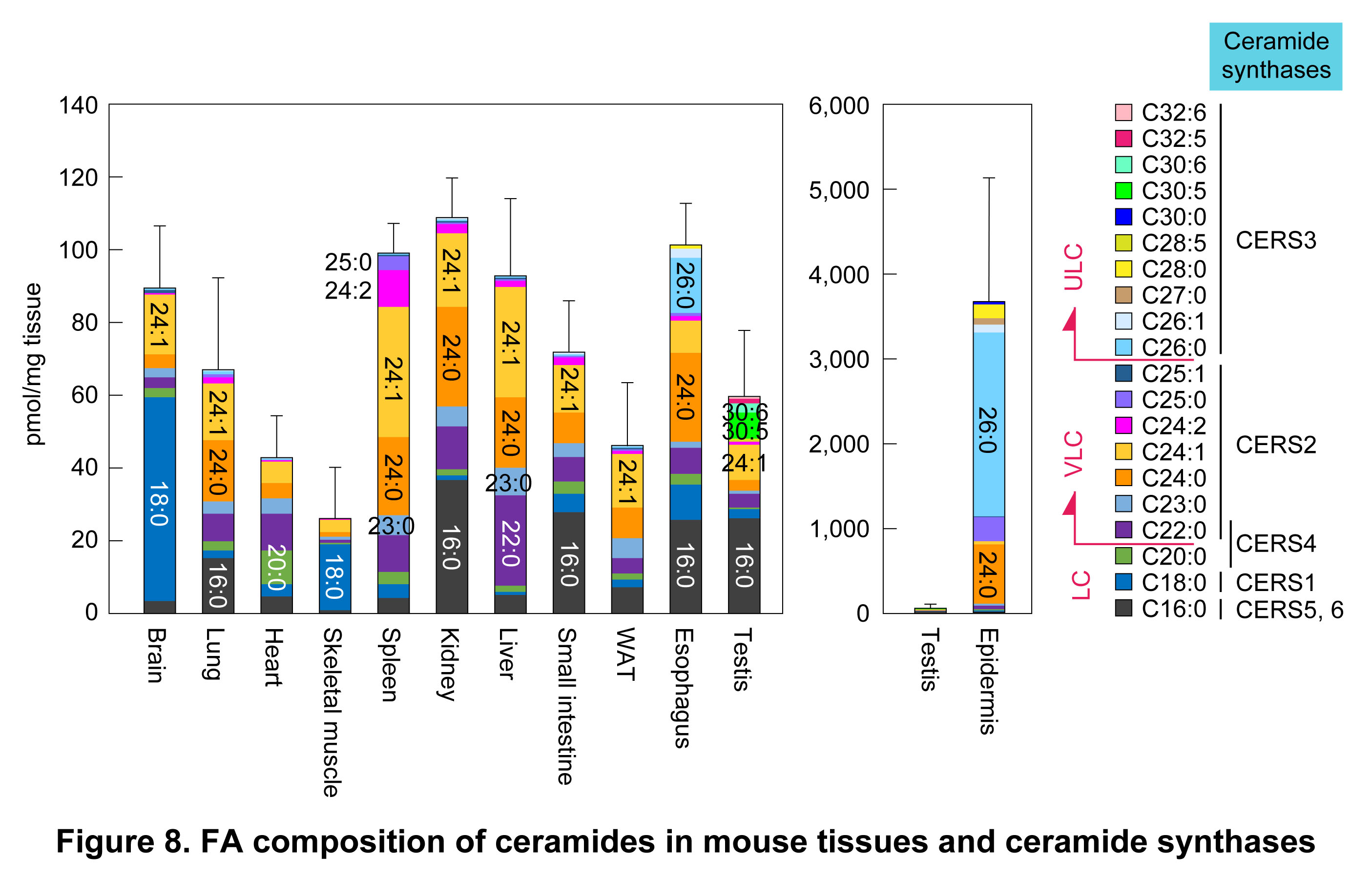
Odd-chain FAs such as C23:0 and C25:0 are relatively abundant in ceramides (Figure 8). These FAs are generated through α-oxidation of 2-hydroxy FAs. For example, 2-hydroxy C24:0 FA is converted to (non-hydroxy) C23:0 FA by α-oxidation, and the resulting C23:0 FA is incorporated into ceramides either directly or after elongation to C25:08, 9). Our laboratory has elucidated the molecular basis of α-oxidation; for further details, see “2. Metabolism of long-chain bases and elucidation of the FA α-oxidation pathway.” Tissue-specific FAs in ceramides include, in addition to the above-mentioned C26:0, testis-specific C30:5 and C30:6. Ceramides and SMs containing these polyunsaturated ULCFAs are essential for spermatogenesis.
FA elongation cycle
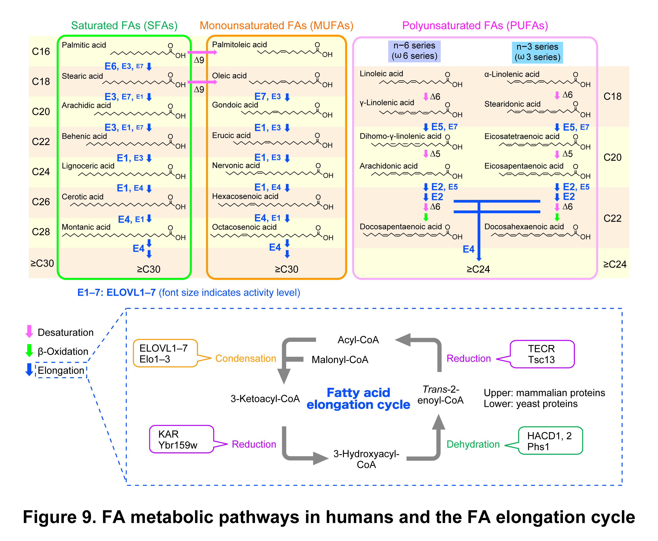
FA elongation occurs in the endoplasmic reticulum. After conversion to acyl-CoA, FAs are elongated by two carbon units per cycle through the FA elongation cycle, which consists of four sequential reactions: condensation, reduction, dehydration, and reduction (Figure 9)1, 2). Among these reactions, the initial condensation reaction is the rate-limiting step and is catalyzed by FA elongases. Mammals possess seven FA elongases (ELOVL1–7), each exhibiting distinct substrate specificities3). Our laboratory systematically analyzed the substrate specificities of ELOVLs using various acyl-CoA substrates in vitro3). This analysis revealed that, among ELOVLs, ELOVL1 exhibits the highest activity for the production of C24 FAs that are characteristic components of sphingolipids. Moreover, knockdown of ELOVL1 in HeLa cells resulted in a decrease in C24 sphingolipids and a concomitant increase in C16 sphingolipids. These changes were associated with altered cellular signaling, including reduced activation of the Src-family kinase Lyn and enhanced apoptosis3, 10). We then generated and analyzed Elovl1 KO mice to investigate the physiological roles of Elovl1 in vivo. These KO mice exhibited neonatal lethality due to impaired skin barrier function4). This phenotype is associated with a reduction in ULCFA-containing ceramides (including acylceramides and protein-bound ceramides) in the epidermis. Decreased levels of VLCFA-containing sphingolipids were also observed in non-epidermal tissues such as the brain and liver, although the affected FA species differed, with reductions in C22- and C24-containing species.
Ceramide synthases corresponding to each FA species are shown on the right side of Figure 8. Mammals possess six ceramide synthases (CERS1–6), each exhibiting distinct substrate specificities toward acyl-CoAs11–13). Thus, individual ceramide species are generated through sequential reactions, in which acyl-CoAs are elongated by specific ELOVL enzymes and subsequently converted into ceramides by specific CERS enzymes via amide bond formation with long-chain bases. The FA composition of ceramides varies among tissues and organs (Figure 8), reflecting differences in the combinations of ELOVL and CERS isozymes expressed. For example, C18:0 FA-containing ceramides (C18:0 ceramides) abundant in the brain are produced by ELOVL6 and CERS1, whereas C24:0 and C24:1 ceramides, widely found across tissues, are generated by ELOVL1 and CERS2. In the epidermis, C26:0 ceramides are synthesized by ELOVL1 and CERS3, while ≥C28 ceramides are produced by ELOVL4 and CERS314). The final chain length of acyl-CoAs produced by ELOVL1 depends on the partnering CERS isozymes: elongation proceeds up to C24 in cooperation with CERS2, whereas it extends to C26 in cooperation with CERS34, 15). C26 acyl-CoA generated by ELOVL1 is further elongated by ELOVL4 to produce longer-chain acyl-CoAs, which serve as substrates for CERS315).
Expression of CERS3 is largely restricted to the epidermis and testis12, 16). In the epidermis, expression of CERS3 and ELOVL4 is induced from the stratum spinosum upward, whereas CERS2 expression decreases and ELOVL1 expression is upregulated in the stratum granulosum4). These changes in expression give rise to the characteristic FA composition of ceramides in the epidermis. We have found that a regulatory mechanism exists between ELOVL and CERS enzymes that function sequentially in ceramide synthesis, in which CERS modulates ELOVL activity. For example, the activities of ELOVL1 and ELOVL4 are enhanced in the presence of CERS2 and CERS3, respectively3, 17). However, the detailed molecular mechanism underlying this regulation remains unclear. Regarding the regulation of CERS activity, we have shown that all CERS isozymes, except for CERS1, are regulated by phosphorylation13). Non-phosphorylatable mutants of CERS2–6 exhibit lower activity than the corresponding wild-type enzymes. CERS2–6 are constitutively phosphorylated under normal growth conditions; however, the mechanisms and conditions regulating their phosphorylation and dephosphorylation remain unclear.
Since whole-body Elovl1 knockout (KO) mice exhibit neonatal lethality due to impaired skin barrier formation4), the roles of VLCFAs in non-epidermal tissues could not be elucidated. To circumvent this limitation, we generated Elovl1 KO mice in which Elovl1 expression was restored specifically in the epidermis. These mice were produced by crossing Elovl1 KO mice with transgenic (Tg) mice expressing Elovl1 under the control of the epidermis-specific involucrin (Inv) promoter. Hereafter, these mice are referred to as Tg-Elovl1 KO mice. Tg-Elovl1 KO mice did not exhibit skin barrier abnormalities and survived to adulthood; however, they developed dry eye, which progressed to corneal opacity after 5 months of age5). For further details, see “5. Prevention of dry eye by meibum lipids.” In addition, Tg-Elovl1 KO mice displayed motor dysfunction and hypomyelination18). In the brains of these mice, levels of galactosylceramides, which are essential for myelin function and structural integrity, were reduced. Shortening of FA chain lengths was also observed in complex sphingolipids such as galactosylceramides, sulfatides, and SMs. The skin and neurological abnormalities observed in whole-body Elovl1 KO and Tg-Elovl1 KO mice were later found to also occur in humans. In 2019, in collaboration with researchers in Germany, we first reported that mutations in ELOVL1 cause a neurocutaneous disorder (IKSHD syndrome) characterized by ichthyotic keratoderma, spasticity, hypomyelination, and dysmorphic facies19).
Peroxisomal β-oxidation is responsible for the degradation of VLCFAs. Transport of VLC acyl-CoAs into peroxisomes is mediated by the ABC transporter ABCD1. Mutations in this gene lead to the accumulation of VLCFAs and cause X-linked adrenoleukodystrophy (X-ALD). Lorenzo’s oil is a mixture (approximately 4:1) of glyceryl trioleate and glyceryl trierucate, which are triacylglycerols of oleic acid (C18:1) and erucic acid (C22:1), respectively, and has been reported to reduce levels of saturated VLCFAs (C24:0 and C26:0) in plasma. However, the mechanism underlying its effect had long been unclear. We demonstrated that a 4:1 mixture of free oleic and erucic acids (corresponding to the hydrolyzed form of Lorenzo’s oil) suppresses the production of saturated VLCFAs in cells by inhibiting ELOVL1 activity20). In contrast, treatment with this FA mixture increased the production of monounsaturated VLCFAs, including C22:1 and C24:1. Although ELOVL1 produces both saturated and monounsaturated VLCFAs in vitro, its contribution in cells and tissues is high for saturated VLCFAs but relatively low for monounsaturated VLCFAs, as monounsaturated VLCFA production receives substantial contributions from other ELOVLs, such as ELOVL3 and ELOVL73). In addition, reduction in ELOVL1 expression, and likely also ELOVL1 activity, induces compensatory upregulation of ELOVL3 and ELOVL7 in various tissues5, 21). Together, these mechanisms may account for the increased production of monounsaturated VLCFAs upon treatment with the FA mixture. Despite the limitations of Lorenzo’s oil in therapeutic efficacy, our findings suggest that ELOVL1 is a promising molecular target for the treatment of X-ALD, and more selective and effective ELOVL1 inhibitors are expected to be developed.
ELOVL4 is responsible for the production of ≥C28 ULCFAs. Lipids containing these FAs are present in limited tissues and organs, including the epidermis (e.g., acylceramides), brain and retina (phosphatidylcholines), sperm (ceramides and SMs), and the meibomian and sebaceous glands (e.g., cholesteryl esters and wax esters)1, 2). Reduced ELOVL4 function causes functional and structural abnormalities in these tissues, leading to macular degeneration, neurological disorders, and ichthyosis. Mutations in ELOVL4 cause three major types of disorders: Stargardt disease 3, spinocerebellar ataxia (SCA) 34, and ichthyosis with spastic quadriplegia and mental retardation (ISQMR), which are associated with truncating mutations with C-terminal deletions, missense mutations with altered function, and loss-of-function mutations, respectively. Among these, SCA34 is an autosomal dominant disorder characterized by cerebellar atrophy and ataxia. Among these, SCA34 is an autosomal dominant disorder characterized by cerebellar atrophy and ataxia. Using neurons differentiated from ES cells carrying SCA34-associated ELOVL4 mutations, we found that ULC PUFA-containing phosphatidylcholines are replaced with shorter species compared to those in wild-type cells22). These results suggest that incomplete elongation of ULC PUFAs by mutant proteins leads to alterations in ULC PUFA-containing phosphatidylcholine composition, which may contribute to the pathogenesis of SCA34.
ELOVL proteins are integral membrane proteins and therefore require solubilization with nonionic detergents, such as Triton X-100, for purification. However, under such solubilized conditions, ELOVLs do not exhibit enzymatic activity. To overcome this limitation, we purified solubilized ELOVL7 and reconstituted it into proteoliposomes, thereby enabling measurement of its enzymatic activity23). The substrate specificity of ELOVL7 determined using this system was consistent with that observed in assays using overexpressed membranes3) and showed high activity toward C18–C20 acyl-CoAs23). We further analyzed ELOVL6, which exhibits activity toward C16:0-CoA, using a similar proteoliposome reconstitution system. When KAR, which catalyzes the second step in the FA elongation cycle, was co-reconstituted with ELOVL6 into proteoliposomes, its activity was enhanced compared to that of ELOVL6 alone24). These results indicate that the first and second steps of the FA elongation cycle are functionally coupled.
Our laboratory identified HACD1–4 as 3-hydroxyacyl-CoA dehydratases that exhibit activity toward 3-hydroxypalmitoyl-CoA in vitro25). In contrast, analyses using cells in which HACD genes were knocked out demonstrated that HACD1 and HACD2 were involved in a wide range of FA elongation reactions, whereas the involvement of HACD3 and HACD4 was minimal or hardly detectable at the cellular level. HACD2 is expressed across diverse tissues and organs and functions as a major enzyme in the FA elongation cycle, whereas HACD1 is specifically expressed and functions in muscle tissues, such as the heart and skeletal muscle. In collaboration with researchers in Israel, we first reported that mutations in HACD1 cause myopathy in humans26). Consistent with this, Hacd1 KO mice also exhibit a myopathic phenotype27).
A missense mutation in TECR (p.P182L), which catalyzes the fourth reaction in the FA elongation cycle, is known to cause non-syndromic intellectual disability. Our laboratory demonstrated that this mutation reduces protein stability and enzymatic activity, and that levels of C24 VLCFA-containing sphingolipids are decreased in patient-derived B-lymphoblastoid cell lines28). Furthermore, reduced levels of TECR led to decreased HACD activity, which catalyzes the third reaction in the FA elongation cycle. Thus, in addition to the first and second reactions, the third and fourth reactions of the FA elongation cycle are also functionally coupled. The TECR P182L mutant protein retains partial activity, and its effects on the FA elongation cycle are relatively modest. Nevertheless, neurological abnormalities are observed, suggesting that VLCFAs play a particularly important role in the nervous system compared to other tissues. Myelin ensheaths axons of neurons and acts as an electrical insulator. It is rich in lipids, and its insulating properties are largely conferred by these lipids. Among these, VLCFA-containing sphingolipids, such as galactosylceramides and sulfatides with C24 FA chains, are essential for myelin formation and maintenance. The TECR p.P182L mutation is therefore likely to impair myelin function through the reduction of these lipid levels. As discussed below, VLCFAs may stabilize highly curved membrane structures such as myelin.
VLCFAs are widely present in eukaryotes, from yeast to humans. In the budding yeast Saccharomyces cerevisiae, VLCFAs are almost exclusively C26 and are found in sphingolipids. Impairment of VLCFA synthesis is lethal in yeast. Enzymes responsible for the four reactions of the FA elongation cycle are conserved among eukaryotes, and yeast Phs1 is a homolog of mammalian HACD1–4 (Figure 9). Our laboratory determined residues essential for Phs1 activity and gained insights into its reaction mechanism29, 30). To investigate the cellular functions of VLCFAs, we performed genetic analyses in yeast. Reduction in VLCFA production causes temperature-sensitive growth in yeast. Taking advantage of this phenotype, we screened for multicopy suppressors that rescue the temperature-sensitive growth defect and identified VPS21, which is involved in vesicular trafficking via the late endosome, also known as the multivesicular body (MVB)31). These findings suggest that sphingolipids containing VLCFAs play an important role in vesicular trafficking via the MVB. Due to their length, VLCFAs are thought to span both leaflets of the lipid bilayer, thereby stabilizing highly curved and otherwise unstable membrane structures. In the MVB, such highly curved membrane structures are formed during intraluminal vesicle formation, which likely underlies the high requirement for VLCFAs in this process. Myelin forms a multilamellar structure surrounding axons and represents a similarly highly curved membrane structure. VLCFA-containing sphingolipids are therefore likely to contribute to the stabilization of myelin, and impairment of VLCFA production may affect the nervous system, as evidenced by neurological disorders caused by mutations in TECR (non-syndromic intellectual disability) and ELOVL1 (IKSHD syndrome).
The catalytic residues and reaction mechanism of trans-2-enoyl-CoA reductase (yeast Tsc13 and mammalian TECR), which catalyzes the fourth reaction in the FA elongation cycle, had remained unclear. Our laboratory demonstrated that Tyr256 in Tsc13 and Tyr248 in TECR function as catalytic residues and proposed a reaction model in which hydride transfer from NADPH generates an enolate intermediate, followed by proton donation from the catalytic tyrosine to complete the reaction32). Furthermore, we revealed interactions between Tsc13 and FA elongases (Elo2 and Elo3), which catalyze the first reaction in the FA elongation cycle in yeast, and showed that these interactions are required to maintain full elongase activity32). These findings indicate that, in addition to the coupling of the first and second reactions and that of the third and fourth reactions, the fourth reaction is also functionally coupled with the first reaction of the subsequent FA elongation cycle.
In summary, the FA elongation cycle proceeds through coordinated coupling among individual reactions, thereby ensuring precise control of VLCFA production and chain length. VLCFAs serve as key components that support the formation and functional maintenance of biological membrane structures. The findings described in this section provide important insights into the molecular mechanisms by which abnormalities in VLCFA metabolism lead to the development of diverse disorders, particularly those affecting the skin and nervous system.
References
- Kihara A. (2012) Very long-chain fatty acids: elongation, physiology and related disorders. J Biochem, 152, 387–395, Review.
- Sassa T, Kihara A. (2014) Metabolism of very long-chain fatty acids: genes and pathophysiology. Biomol Ther, 22, 83–92, Review.
- Ohno Y, Suto S, Yamanaka M, Mizutani Y, Mitsutake S, Igarashi Y, Sassa T, Kihara A. (2010) ELOVL1 production of C24 acyl-CoAs is linked to C24 sphingolipid synthesis. Proc Natl Acad Sci USA, 107, 18439–18444.
- Sassa T, Ohno Y, Suzuki S, Nomura T, Nishioka C, Kashiwagi T, Hirayama T, Akiyama M, Taguchi R, Shimizu H, Itohara S, Kihara A. (2013) Impaired epidermal permeability barrier in mice lacking Elovl1, the gene responsible for very-long-chain fatty acid production. Mol Cell Biol, 33, 2787–2796.
- Sassa T, Tadaki M, Kiyonari H, Kihara A. (2018) Very long-chain tear film lipids produced by fatty acid elongase ELOVL1 prevent dry eye disease in mice. FASEB J, 32, 2966–2978.
- Sugimoto M, Shimizu Y, Yoshioka T, Wakabayashi M, Tanaka Y, Higashino K, Numata Y, Sakai S, Kihara A, Igarashi Y, Kuge Y. (2015) Histological analyses by matrix-assisted laser desorption/ionization-imaging mass spectrometry reveal differential localization of sphingomyelin molecular species regulated by particular ceramide synthase in mouse brains. Biochim Biophys Acta, 1851, 1554–1565.
- Edagawa M, Sawai M, Ohno Y, Kihara A. (2018) Widespread tissue distribution and synthetic pathway of polyunsaturated C24:2 sphingolipids in mammals. Biochim Biophys Acta Mol Cell Biol Lipids, 1863, 1441–1448.
- Mori K, Obara T, Seki N, Miyamoto M, Naganuma T, Kitamura T, Kihara A. (2020) Catalytic residues, substrate specificity, and role in carbon starvation of the 2-hydroxy FA dioxygenase Mpo1 in yeast. J Lipid Res, 61, 1104–1114.
- Mori K, Naganuma T, Kihara A. (2023) Role of 2-hydroxy acyl-CoA lyase HACL2 in odd-chain fatty acid production via α-oxidation in vivo. Mol Biol Cell, 34, ar85.
- Sassa T, Suto S, Okayasu Y, Kihara A. (2012) A shift in sphingolipid composition from C24 to C16 increases susceptibility to apoptosis in HeLa cells. Biochim Biophys Acta, 1821, 1031–1037.
- Mizutani Y, Kihara A, Igarashi Y. (2005) Mammalian Lass6 and its related family members regulate synthesis of specific ceramides. Biochem J, 390, 263–271.
- Mizutani Y, Kihara A, Igarashi Y. (2006) LASS3 (longevity assurance homologue 3) is a mainly testis-specific (dihydro)ceramide synthase with relatively broad substrate specificity. Biochem J, 398, 531–538.
- Sassa T, Hirayama T, Kihara A. (2016) Enzyme activities of the ceramide synthases CERS2–6 are regulated by phosphorylation in the C-terminal region. J Biol Chem, 291, 7477–7487.
- Kihara A. (2016) Synthesis and degradation pathways, functions, and pathology of ceramides and epidermal acylceramides. Prog Lipid Res, 63, 50–69, Review.
- Ohno Y, Nakamichi S, Ohkuni A, Kamiyama N, Naoe A, Tsujimura H, Yokose U, Sugiura K, Ishikawa J, Akiyama M, Kihara A. (2015) Essential role of the cytochrome P450 CYP4F22 in the production of acylceramide, the key lipid for skin permeability barrier formation. Proc Natl Acad Sci USA, 112, 7707–7712.
- Mizutani Y, Kihara A, Chiba H, Tojo H, Igarashi Y. (2008) 2-Hydroxy-ceramide synthesis by ceramide synthase family: enzymatic basis for the preference of FA chain length. J Lipid Res, 49, 2356–2364.
- Mizutani Y, Sun H, Ohno Y, Sassa T, Wakashima T, Obara M, Yuyama K, Kihara A, Igarashi Y. (2013) Cooperative synthesis of ultra long-chain fatty acid and ceramide during keratinocyte differentiation. PLoS One, 8, e67317.
- Isokawa M, Sassa T, Hattori S, Miyakawa T, Kihara A. (2019) Reduced chain length in myelin sphingolipids and poorer motor coordination in mice deficient in the fatty acid elongase Elovl1. FASEB BioAdvances, 1, 747–759.
- Mueller N, Sassa T, Morales-Gonzalez S, Schneider J, Salchow D J, Seelow D, Knierim E, Stenzel W, Kihara A, Schuelke M. (2019) De novo mutation in ELOVL1 causes ichthyosis, acanthosis nigricans, hypomyelination, spastic paraplegia, high frequency deafness and optic atrophy. J Med Genet, 56, 164–175.
- Sassa T, Wakashima T, Ohno Y, Kihara A. (2014) Lorenzo’s oil inhibits ELOVL1 and lowers the level of sphingomyelin with a saturated very long-chain fatty acid. J Lipid Res, 55, 524–530.
- Hiranuma T, Sassa T, Kihara A. (2025) Relationship between time-dependent epidermal ceramide composition changes and skin barrier function in adult mice. Mol Biol Cell, 36, ar57.
- Tamura Y, Sassa T, Nishizawa T, Kihara A. (2023) Incomplete elongation of ultra-long-chain polyunsaturated acyl-CoAs by the fatty acid elongase ELOVL4 in spinocerebellar ataxia type 34. Mol Cell Biol, 43, 85–101.
- Naganuma T, Sato Y, Sassa T, Ohno Y, Kihara A. (2011) Biochemical characterization of the very long-chain fatty acid elongase ELOVL7. FEBS Lett, 585, 3337–3341.
- Naganuma T, Kihara A. (2014) Two modes of regulation of the fatty acid elongase ELOVL6 by the 3-ketoacyl-CoA reductase KAR in the fatty acid elongation cycle. PLoS One, 9, e101823.
- Ikeda M, Kanao Y, Yamanaka M, Sakuraba H, Mizutani Y, Igarashi Y, Kihara A. (2008) Characterization of four mammalian 3-hydroxyacyl-CoA dehydratases involved in very long-chain fatty acid synthesis. FEBS Lett, 582, 2435–2440.
- Muhammad E, Reish O, Ohno Y, Scheetz T, Deluca A, Searby C, Regev M, Benyamini L, Fellig Y, Kihara A, Sheffield V C, Parvari R. (2013) Congenital myopathy is caused by mutation of HACD1. Hum Mol Genet, 22, 5229–5236.
- Blondelle J, Ohno Y, Gache V, Guyot S, Storck S, Blanchard-Gutton N, Barthelemy I, Walmsley G, Rahier A, Gadin S, Maurer M, Guillaud L, Prola A, Ferry A, Aubin-Houzelstein G, Demarquoy J, Relaix F, Piercy R J, Blot S, Kihara A, Tiret L, Pilot-Storck F. (2015) HACD1, a regulator of membrane composition and fluidity, promotes myoblast fusion and skeletal muscle growth. J Mol Cell Biol, 7, 429–440.
- Abe K, Ohno Y, Sassa T, Taguchi R, Caliskan M, Ober C, Kihara A. (2013) Mutation for nonsyndromic mental retardation in the trans-2-enoyl-CoA reductase TER gene involved in fatty acid elongation impairs the enzyme activity and stability, leading to change in sphingolipid profile. J Biol Chem, 288, 36741–36749.
- Kihara A, Sakuraba H, Ikeda M, Denpoh A, Igarashi Y. (2008) Membrane topology and essential amino acid residues of Phs1, a 3-hydroxyacyl-CoA dehydratase involved in very long-chain fatty acid elongation. J Biol Chem, 283, 11199–11209.
- Yazawa T, Naganuma T, Yamagata M, Kihara A. (2013) Identification of residues important for the catalysis, structure maintenance, and substrate specificity of yeast 3-hydroxyacyl-CoA dehydratase Phs1. FEBS Lett, 587, 804–809.
- Obara K, Kojima R, Kihara A. (2013) Effects on vesicular transport pathways at the late endosome in cells with limited very long-chain fatty acids. J Lipid Res, 54, 831–842.
- Kato R, Takenaka Y, Ohno Y, Kihara A. (2024) Catalytic mechanism of trans-2-enoyl-CoA reductases in the fatty acid elongation cycle and its cooperative action with fatty acid elongases. J Biol Chem, 300, 105656.